A new Level-set based Protocol for Accurate Bone Segmentation from CT Imaging
Publication
Metrics
AI Quick Summary
This paper proposes a two-step level-set based protocol for accurate bone segmentation from CT imaging, combining user-defined pre-segmentation with fully automatic refinement. The refined segmentation achieves high sub-pixel accuracy validated against a gold standard model, demonstrating effective bone segmentation across different reconstruction protocols.
Paper Preview
Abstract
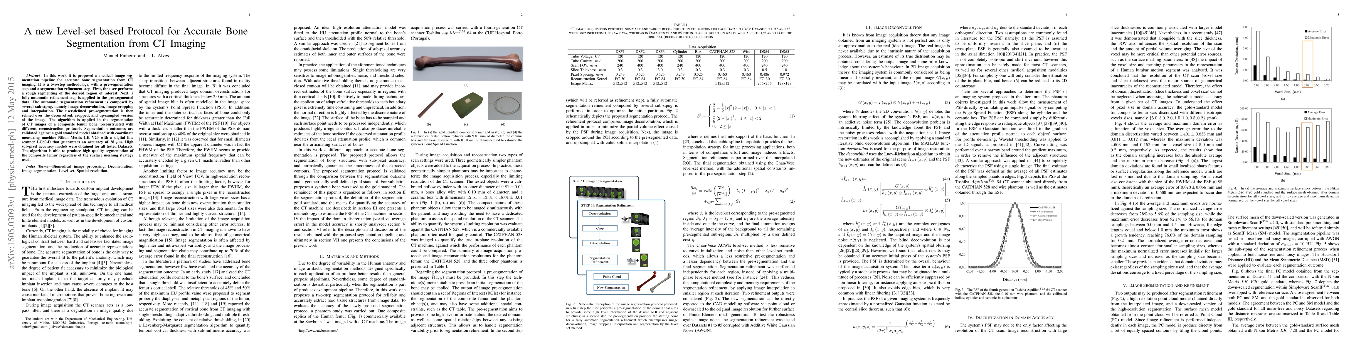
In this work it is proposed a medical image segmentation pipeline for accurate bone segmentation from CT imaging. It is a two-step methodology, with a pre-segmentation step and a segmentation refinement step. First, the user performs a rough segmenting of the desired region of interest. Next, a fully automatic refinement step is applied to the pre-segmented data. The automatic segmentation refinement is composed by several sub-stpng, namely image deconvolution, image cropping and interpolation. The user-defined pre-segmentation is then refined over the deconvolved, cropped, and up-sampled version of the image. The algorithm is applied in the segmentation of CT images of a composite femur bone, reconstructed with different reconstruction protocols. Segmentation outcomes are validated against a gold standard model obtained with coordinate measuring machine Nikon Metris LK V20 with a digital line scanner LC60-D that guarantees an accuracy of 28 $\mu m$. High sub-pixel accuracy models were obtained for all tested Datasets. The algorithm is able to produce high quality segmentation of the composite femur regardless of the surface meshing strategy used.
AI Key Findings
Get AI-generated insights about this paper's methodology, results, significance, and more — seven facets brought into focus.
Impact
Paper Details
PDF Preview
Key Terms
Citation Network
Current paper (gray), citations (green), references (blue)
Display is limited for performance on very large graphs.
Discussion 0