ArxivLens

Quick Summary:

This paper introduces a novel deep learning method combining a 3D V-Net with shape priors to segment left ventricular myocardium in myocardial perfusion SPECT images, achieving high Dice similarity coefficients and low Hausdorff distances, demonstrating its effectiveness for accurate LV function assessment.

DOI: 10.1016/j.compbiomed.2023.106954
arXiv Id: 2206.03603
Comments: 21 pages, 14 figures
Date Published: 2023-12-22
Date Updated: 2023-12-22

Categories:

Key Terms (TF-IDF):

mm (0.258)
priors (0.227)
extracting (0.208)
shape (0.199)
segmentation (0.185)
validate (0.180)
proposed method (0.167)
achieved (0.155)
dp (0.143)
hd (0.137)
hausdorff (0.136)
model training (0.133)
integrates (0.129)
high accuracy (0.128)
proposed (0.128)
guided (0.124)
conclusion (0.122)
ground truth (0.120)
new method (0.120)
method (0.119)
License: http://creativecommons.org/licenses/by/4.0/

Summary:

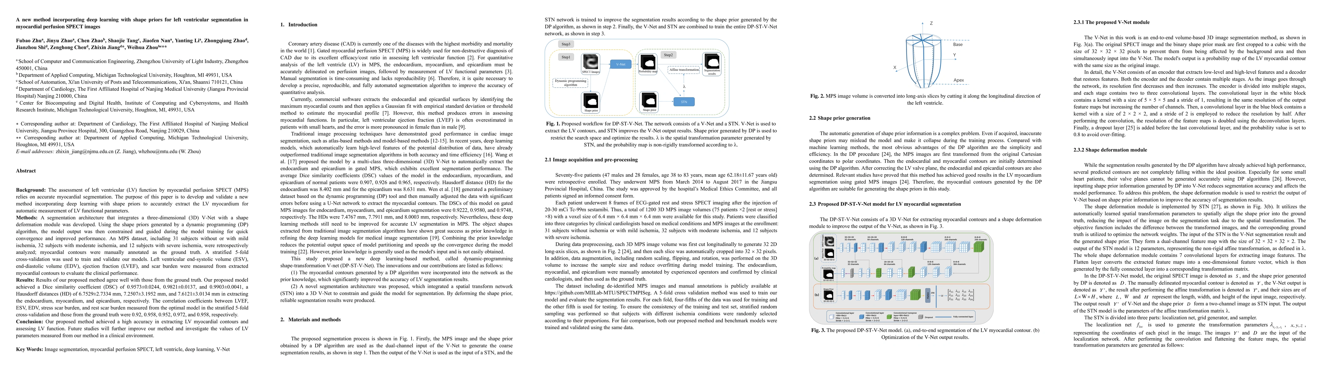
Background: The assessment of left ventricular (LV) function by myocardial perfusion SPECT (MPS) relies on accurate myocardial segmentation. The purpose of this paper is to develop and validate a new method incorporating deep learning with shape priors to accurately extract the LV myocardium for automatic measurement of LV functional parameters. Methods: A segmentation architecture that integrates a three-dimensional (3D) V-Net with a shape deformation module was developed. Using the shape priors generated by a dynamic programming (DP) algorithm, the model output was then constrained and guided during the model training for quick convergence and improved performance. A stratified 5-fold cross-validation was used to train and validate our models. Results: Results of our proposed method agree well with those from the ground truth. Our proposed model achieved a Dice similarity coefficient (DSC) of 0.9573(0.0244), 0.9821(0.0137), and 0.9903(0.0041), a Hausdorff distances (HD) of 6.7529(2.7334) mm, 7.2507(3.1952) mm, and 7.6121(3.0134) mm in extracting the endocardium, myocardium, and epicardium, respectively. Conclusion: Our proposed method achieved a high accuracy in extracting LV myocardial contours and assessing LV function.

Citation & Reference Network Visualization

This graph shows how this paper is connected to other research. The current paper is shown in dark gray, papers that cite it are in green, and papers it references are in blue. You can:

Current Paper
Papers citing this work (Citations)
Papers cited by this work (References)

• Hover over any node to see the paper title

• Click a node to view that paper's details

• Drag nodes to rearrange the graph

• Use the zoom controls (bottom right) to zoom in/out

Citations and References

Title Authors Year Actions
Myocardial perfusion imaging SPECT left ventricle segmentation with graphs
Ádám István Szűcs, Béla Kári, Oszkár Pártos
2025
View Paper
A Deep Learning Approach for Robust Segmentation of Left Ventricular Wall in Cardiac SPECT Images
E. Arin, Ş. İ. Yetik, J. Brankov, Y. Yang, M. King
2024
View Paper
Automatic reorientation to generate short-axis myocardial PET images
Yuling Yang, Fanghu Wang, Xu Han, Hui Xu, Yangmei Zhang, Weiping Xu, Shuxia Wang, Lijun Lu
2024
View Paper
Multi-modality deep learning-based [68Ga]Ga-DOTA-FAPI-04 PET polar map generation: potential value in detecting reactive fibrosis after myocardial infarction.
Xiaoya Qiao, Hanzhong Wang, Hongping Meng, Yun Xi, David Dagan Feng, Biao Li, Xiaoxiang Yan, Min Zhang, Qiu Huang
2024
View Paper
Unfolding Explainable AI for Brain Tumor Segmentation
Muhammad Hassan, A. A. Fateh, Jieqiong Lin, Yijiang Zhuang, Guisen Lin, Hairui Xiong, You Zhou, Peiwu Qin, Hongwu Zeng
2024
View Paper
Deep Learning Artificial Intelligence for Single Photon Emission Computed Tomography: A Comprehensive Overview and Future Perspectives
Rongyu Cui, Xin Xie, Long Fan, Xiaoli Yang, Yixuan Ren, Weihai Qin
2024
View Paper
Continuous Max-Flow Augmentation of Self-Supervised Few-Shot Learning on Spect Left Ventricles
Ádám István Szücs, Béla Kári, Oszkár Pártos
2024
View Paper
Multi-residual 2D network integrating spatial correlation for whole heart segmentation
Yan Huang, Jinzhu Yang, Qi Sun, Yuliang Yuan, Honghe Li, Yang Hou
2024
View Paper
Novel Method for Measuring a Wear Scar Using Deep Learning
Sebin Lee, Taewan Kim, Seungchul Lee, Sung-Ho Hong
2023
View Paper
Self-supervised segmentation of myocardial perfusion imaging SPECT left ventricles
Ádám István Szűcs, Kristóf Horváth, Katalin Sólyomvári, Ádám Zlehovszky, Béla Kári
2023
View Paper
3D fusion between SPECT myocardial perfusion imaging and invasive coronary angiography to guide the treatment for patients with stable CAD
Z. Xu, S. Malhotra, C. Zhao, J. Jiang, A. Vij, Z. Ye, R. Hua, C. Li, C. Wang, W. Zhou
2023
View Paper
Automatic reorientation by deep learning to generate short-axis SPECT myocardial perfusion images
Fubao Zhu, Guojie Wang, Chen Zhao, S. Malhotra, Min Zhao, Zhuo He, Jianzhou Shi, Zhixin Jiang, Weihua Zhou
2022
View Paper
结合频域先验和特征增强的心脏图像分割方法
陈柯炎 Chen Keyan, 刘巧红 Liu Qiaohong, 韩啸翔 Han Xiaoxiang, 林元杰 Lin Yuanjie, 张维坤 Zhang Weikun
2024
View Paper
Computer Vision-based Efficient Segmentation Method for Left Ventricular Epicardium and Endocardium using Deep Learning
Saifuddin Saif, Trung Duong, Zachary Holden
2023
View Paper

Comments (0)

This website uses cookies to ensure you get the best experience. Learn more