A New Path to Nanoscale Cellular Analysis with Monochromated Electron Energy-Loss Spectroscopy
Publication
Metrics
AI Quick Summary
This paper introduces a novel method for nanoscale compositional analysis of biological materials using monochromated electron energy-loss spectroscopy (EELS) in a scanning transmission electron microscope. The study successfully maps the chemical composition of a cucumber stem's vascular system, revealing distinct physical and vibrational signatures from different cellular regions, and identifies the compositional origins of these signatures through combined spectroscopic and first-principles calculations.
Paper Preview
Abstract
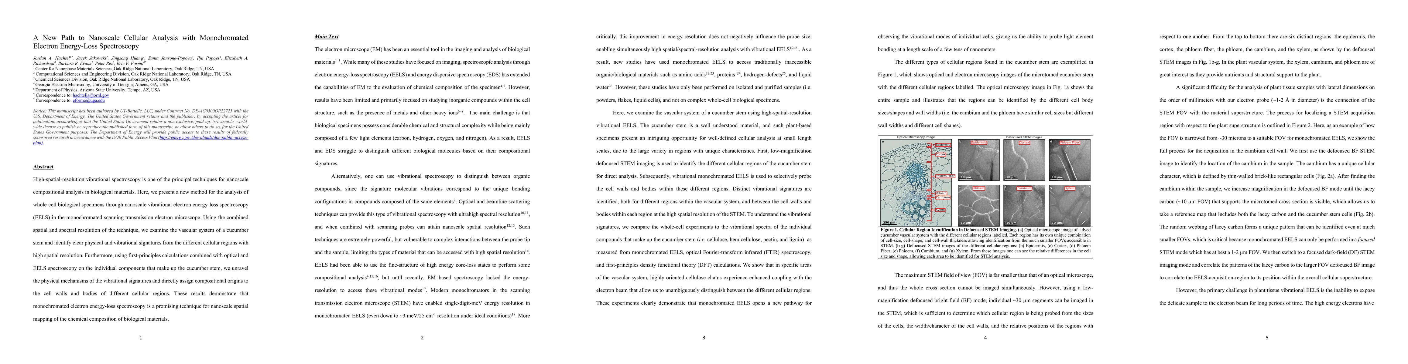
High-spatial-resolution vibrational spectroscopy is one of the principal techniques for nanoscale compositional analysis in biological materials. Here, we present a new method for the analysis of whole-cell biological specimens through nanoscale vibrational electron energy-loss spectroscopy (EELS) in the monochromated scanning transmission electron microscope. Using the combined spatial and spectral resolution of the technique, we examine the vascular system of a cucumber stem and identify clear physical and vibrational signatures from the different cellular regions with high spatial resolution. Furthermore, using first-principles calculations combined with optical and EELS spectroscopy on the individual components that make up the cucumber stem, we unravel the physical mechanisms of the vibrational signatures and directly assign compositional origins to the cell walls and bodies of different cellular regions. These results demonstrate that monochromated electron energy-loss spectroscopy is a promising technique for nanoscale spatial mapping of the chemical composition of biological materials.
AI Key Findings
Get AI-generated insights about this paper's methodology, results, significance, and more — seven facets brought into focus.
Impact
Paper Details
Authors
PDF Preview
Key Terms
Citation Network
Current paper (gray), citations (green), references (blue)
Display is limited for performance on very large graphs.
Discussion 0