Publication
Metrics
AI Quick Summary
This study employs a next-generation qPlus sensor-based AFM setup to resolve the crystal-like structures of Pyrobaculum aerophilium S-layer proteins in both air and liquid, overcoming previous challenges with residual detergent visibility. The new liquid cell and tip protocol significantly improved imaging quality compared to TEM.
Paper Preview
Abstract
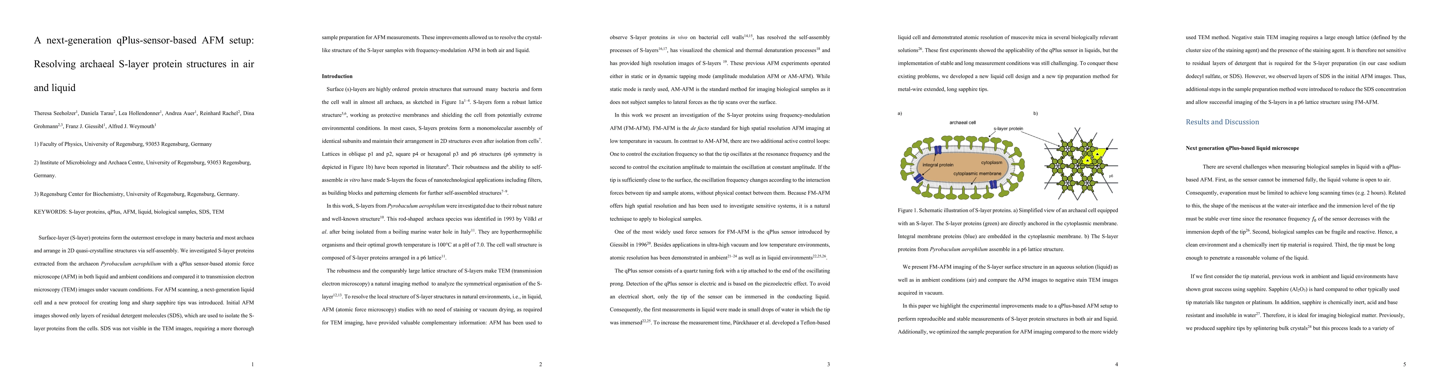
Surface-layer (S-layer) proteins form the outermost envelope in many bacteria and most archaea and arrange in 2D quasi-crystalline structures via self-assembly. We investigated S-layer proteins extracted from the archaeon Pyrobaculum aerophilium with a qPlus sensor-based atomic force microscope (AFM) in both liquid and ambient conditions and compared it to transmission electron microscopy (TEM) images under vacuum conditions. For AFM scanning, a next-generation liquid cell and a new protocol for creating long and sharp sapphire tips was introduced. Initial AFM images showed only layers of residual detergent molecules (SDS), which are used to isolate the S-layer proteins from the cells. SDS was not visible in the TEM images, requiring a more thorough sample preparation for AFM measurements. These improvements allowed us to resolve the crystal-like structure of the S-layer samples with frequency-modulation AFM in both air and liquid.
AI Key Findings
Get AI-generated insights about this paper's methodology, results, significance, and more — seven facets brought into focus.
Impact
Paper Details
Authors
PDF Preview
Key Terms
Citation Network
Current paper (gray), citations (green), references (blue)
Display is limited for performance on very large graphs.
Discussion 0