Summary
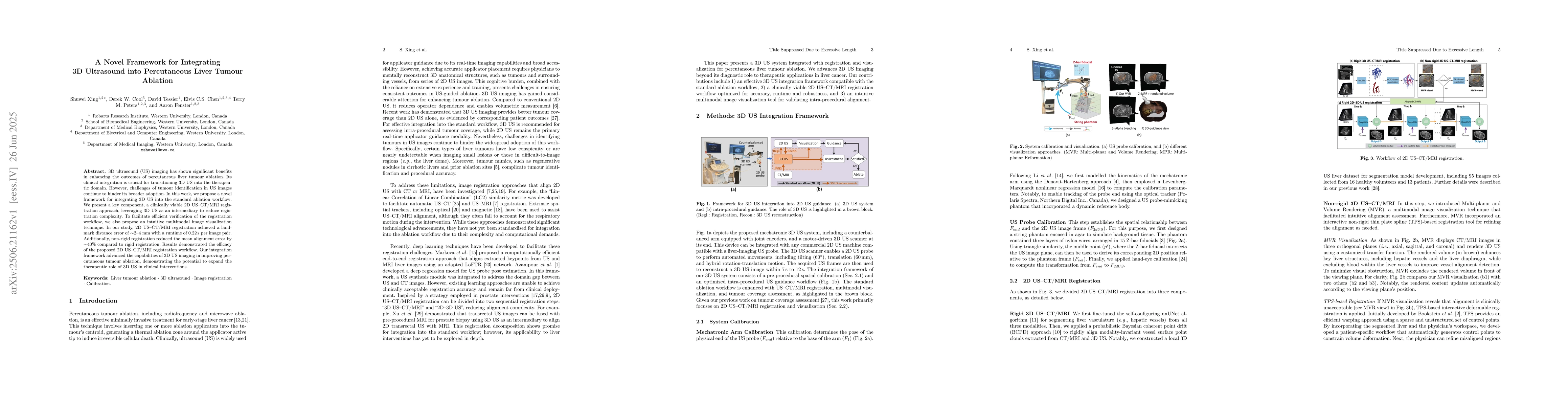
3D ultrasound (US) imaging has shown significant benefits in enhancing the outcomes of percutaneous liver tumour ablation. Its clinical integration is crucial for transitioning 3D US into the therapeutic domain. However, challenges of tumour identification in US images continue to hinder its broader adoption. In this work, we propose a novel framework for integrating 3D US into the standard ablation workflow. We present a key component, a clinically viable 2D US-CT/MRI registration approach, leveraging 3D US as an intermediary to reduce registration complexity. To facilitate efficient verification of the registration workflow, we also propose an intuitive multimodal image visualization technique. In our study, 2D US-CT/MRI registration achieved a landmark distance error of approximately 2-4 mm with a runtime of 0.22s per image pair. Additionally, non-rigid registration reduced the mean alignment error by approximately 40% compared to rigid registration. Results demonstrated the efficacy of the proposed 2D US-CT/MRI registration workflow. Our integration framework advanced the capabilities of 3D US imaging in improving percutaneous tumour ablation, demonstrating the potential to expand the therapeutic role of 3D US in clinical interventions.
AI Key Findings
Generated Jul 01, 2025
Methodology
The study developed a framework integrating 3D ultrasound with CT/MRI for liver tumor ablation, utilizing a 2D US-CT/MRI registration approach, multimodal visualization, and deformable registration techniques, validated through experiments on healthy volunteers and patients.
Key Results
- 2D US-CT/MRI registration achieved a landmark error of approximately 2-4 mm with a runtime of 0.22 seconds per image pair.
- Non-rigid registration reduced the mean alignment error by about 40% compared to rigid registration.
- The registration workflow met clinical accuracy thresholds, with errors less than 5 mm in most cases, demonstrating clinical feasibility.
Significance
This research enhances the integration of 3D US into liver tumor ablation procedures, potentially improving tumor localization, procedural safety, and outcomes in clinical interventions.
Technical Contribution
The work introduces a novel multimodal registration framework leveraging 3D US as an intermediary, combined with intuitive visualization and deformable registration techniques for improved image alignment.
Novelty
This study is distinguished by its clinically viable 2D US-CT/MRI registration approach, integrated multimodal visualization, and comprehensive validation in both healthy and patient cases, advancing the application of 3D US in liver tumor ablation.
Limitations
- The study primarily involved healthy volunteers and a limited number of patient cases, which may not fully represent clinical variability.
- Runtime optimization for real-time application in diverse patient populations remains to be further developed.
Future Work
- Conduct larger-scale patient studies to validate the workflow across diverse clinical scenarios.
- Optimize registration algorithms for faster processing to facilitate real-time intraoperative guidance.
Paper Details
PDF Preview
Citation Network
Current paper (gray), citations (green), references (blue)
Display is limited for performance on very large graphs.
Similar Papers
Found 4 papersDeep Regression 2D-3D Ultrasound Registration for Liver Motion Correction in Focal Tumor Thermal Ablation
Aaron Fenster, Shuwei Xing, Derek W. Cool et al.
Novel Combination of a 1.4mm Radiofrequency Ablation Catheter with Ultrathin Bronchoscope for Subpleural Peripheral Lung Tumour Ablation.
Voon, Pei Jye, Kho, Sze Shyang, Chai, Chan Sin et al.
No citations found for this paper.
Comments (0)