A Practical Reconstruction Method for Three-Dimensional Phase Contrast Atomic Electron Tomography
Publication
Metrics
AI Quick Summary
This paper introduces a novel algorithm for reconstructing 3D electrostatic potentials at atomic resolution using phase contrast imaging in high-resolution transmission electron microscopy. The method effectively handles dynamical and strong phase scattering, achieving accurate results with significantly lower electron doses compared to existing techniques.
Paper Preview
Abstract
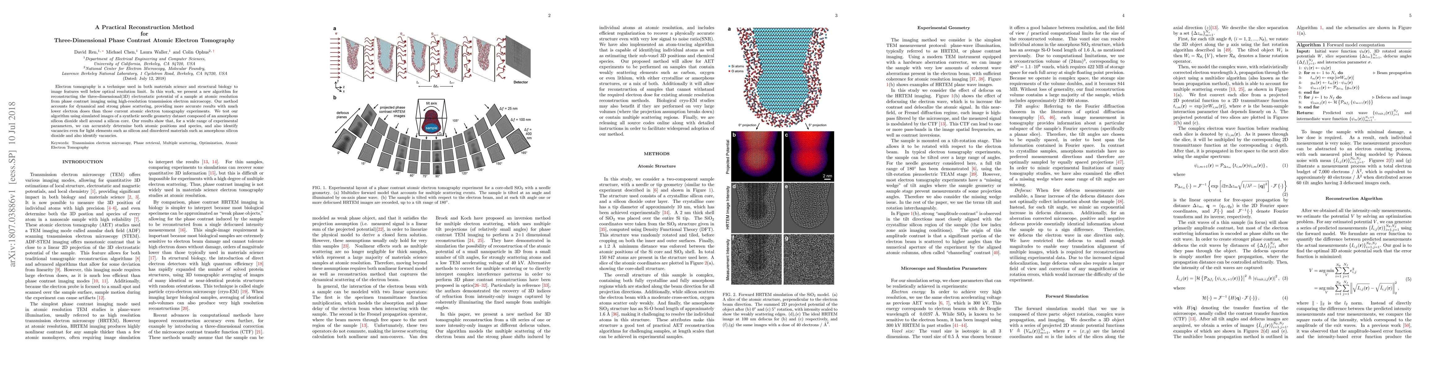
Electron tomography is a technique used in both materials science and structural biology to image features well below optical resolution limit. In this work, we present a new algorithm for reconstructing the three-dimensional(3D) electrostatic potential of a sample at atomic resolution from phase contrast imaging using high-resolution transmission electron microscopy. Our method accounts for dynamical and strong phase scattering, providing more accurate results with much lower electron doses than those current atomic electron tomography experiments. We test our algorithm using simulated images of a synthetic needle geometry dataset composed of an amorphous silicon dioxide shell around a silicon core. Our results show that, for a wide range of experimental parameters, we can accurately determine both atomic positions and species, and also identify vacancies even for light elements such as silicon and disordered materials such as amorphous silicon dioxide and also identify vacancies.
AI Key Findings
Get AI-generated insights about this paper's methodology, results, significance, and more — seven facets brought into focus.
Impact
Paper Details
PDF Preview
Key Terms
Citation Network
Current paper (gray), citations (green), references (blue)
Display is limited for performance on very large graphs.
Discussion 0