01
MethodologyHow they did it
A deep learning-based approach was used to estimate fetal head circumference (HC) from 2D ultrasound images.
This paper reviews recent deep-learning algorithms for fetal ultrasound image analysis, focusing on methodologies and applications in standard-plane detection, anatomical-structure analysis, and biometry parameter estimation. It critically assesses current limitations and challenges, aiming to bridge research and clinical practice.
This paper reviews recent deep-learning algorithms for fetal ultrasound image analysis, focusing on methodologies and applications in standard-plane detection, anatomical-structure analysis, and biometry parameter estimation. It critically assesses current limitations and challenges, aiming to bridge research and clinical practice.
A deep learning-based approach was used to estimate fetal head circumference (HC) from 2D ultrasound images. More in Methodology →
The proposed method achieved a mean absolute error (MAE) of 1.95 mm in estimating HC — The method demonstrated high accuracy in segmenting the fetal head and identifying relevant anatomical structures More in Key Results →
Accurate HC estimation is crucial for monitoring fetal growth and detecting potential complications during pregnancy. More in Significance →
Insufficient amniotic fluid can lead to inaccurate HC measurements — The method may not perform well with images of poor quality or resolution More in Limitations →
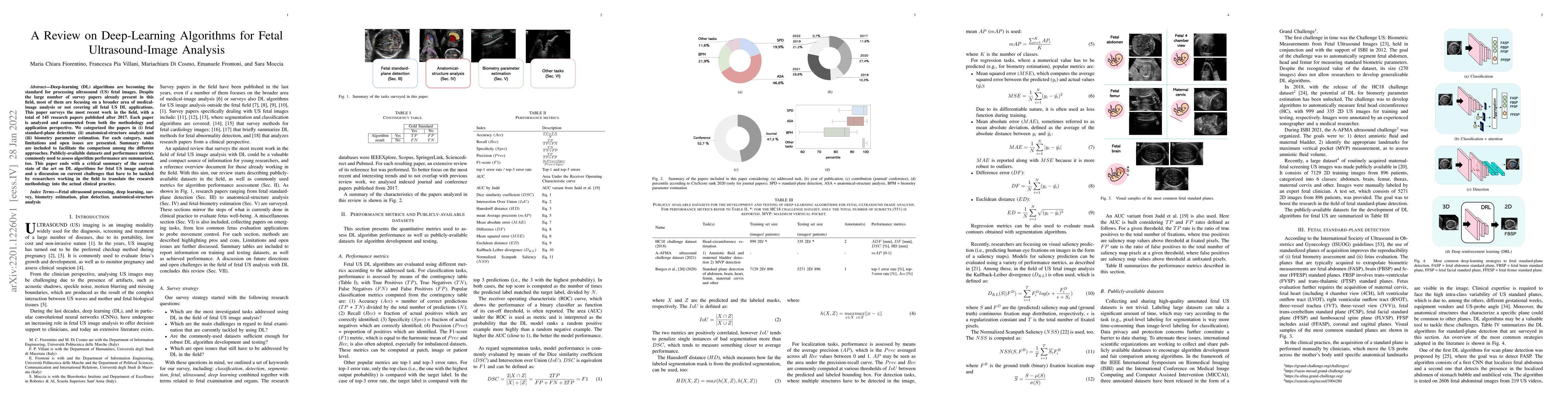
Deep-learning (DL) algorithms are becoming the standard for processing ultrasound (US) fetal images. Despite a large number of survey papers already present in this field, most of them are focusing on a broader area of medical-image analysis or not covering all fetal US DL applications. This paper surveys the most recent work in the field, with a total of 145 research papers published after 2017. Each paper is analyzed and commented on from both the methodology and application perspective. We categorized the papers in (i) fetal standard-plane detection, (ii) anatomical-structure analysis, and (iii) biometry parameter estimation. For each category, main limitations and open issues are presented. Summary tables are included to facilitate the comparison among the different approaches. Publicly-available datasets and performance metrics commonly used to assess algorithm performance are summarized, too. This paper ends with a critical summary of the current state of the art on DL algorithms for fetal US image analysis and a discussion on current challenges that have to be tackled by researchers working in the field to translate the research methodology into the actual clinical practice.
Seven facets of this paper, analysed and brought into focus by AI.
Accurate HC estimation is crucial for monitoring fetal growth and detecting potential complications during pregnancy.
A deep learning-based approach was used to estimate fetal head circumference (HC) from 2D ultrasound images.
Accurate HC estimation is crucial for monitoring fetal growth and detecting potential complications during pregnancy.
The proposed method utilizes a custom-designed CNN architecture and Hough transformation for AC measurement.
The work presents a novel approach to estimating fetal head circumference from 2D ultrasound images using a combination of segmentation and regression techniques
Current paper (gray), citations (green), references (blue)
Display is limited for performance on very large graphs.
Discussion 0