This study employed a mixed-methods approach combining fMRI data analysis with histological examination to investigate the functional organization of the brain.
Publication
Metrics
AI Quick Summary
This paper reviews brain parcellation methods for MRI datasets, categorizing them into anatomical, functional, and structural parcellations based on T1-weighted, fMRI, and DWI datasets respectively. It provides a taxonomy of methods, compares their strengths and weaknesses, and identifies current challenges in brain parcellation research.
Quick Answers
What is "A Review on MR Based Human Brain Parcellation Methods" about?
This paper reviews brain parcellation methods for MRI datasets, categorizing them into anatomical, functional, and structural parcellations based on T1-weighted, fMRI, and DWI datasets respectively. It provides a taxonomy of methods, compares their strengths and weaknesses, and identifies current challenges in brain parcellation research.
What methodology did the authors use?
This study employed a mixed-methods approach combining fMRI data analysis with histological examination to investigate the functional organization of the brain. More in Methodology →
What are the key results?
The number of brain regions identified using functional connectivity profiles was significantly higher than those obtained from resting-state fMRI activity. — Functional parcellations constructed from task-evoked fMRI activity showed greater variability across subjects compared to those constructed from resting-state data. More in Key Results →
Why is this work significant?
This study highlights the importance of considering dynamic changes in brain organization across different brain states and tasks, which can inform our understanding of neurological disorders. More in Significance →
What are the main limitations?
The sample size was relatively small, limiting generalizability to larger populations. — The fMRI data used were collected from a specific population with potential biases that may not be representative of the broader population. More in Limitations →
Abstract
Brain parcellations play a ubiquitous role in the analysis of magnetic resonance imaging (MRI) datasets. Over 100 years of research has been conducted 
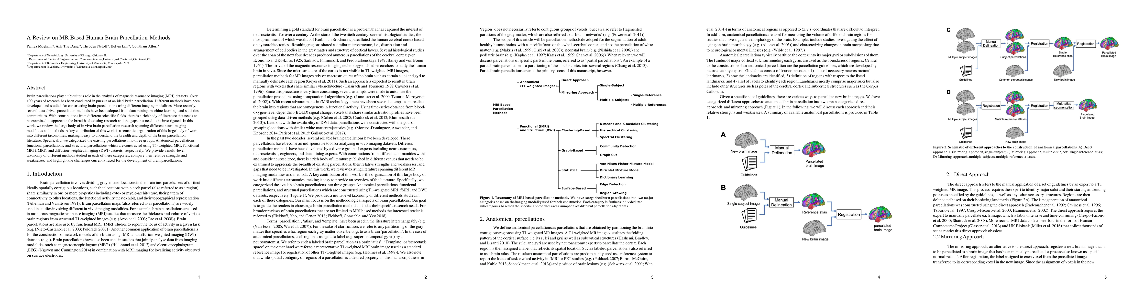
in pursuit of an ideal brain parcellation. Different methods have been developed and studied for constructing brain parcellations using different imaging modalities. More recently, several data-driven parcellation methods have been adopted from data mining, machine learning, and statistics communities. With contributions from different scientific fields, there is a rich body of literature that needs to be examined to appreciate the breadth of existing research and the gaps that need to be investigated. In this work, we review the large body of in vivo brain parcellation research spanning different neuroimaging modalities and methods. A key contribution of this work is a semantic organization of this large body of work into different taxonomies, making it easy to understand the breadth and depth of the brain parcellation literature. Specifically, we categorized the existing parcellations into three groups: Anatomical parcellations, functional parcellations, and structural parcellations which are constructed using T1-weighted MRI, functional MRI (fMRI), and diffusion-weighted imaging (DWI) datasets, respectively. We provide a multi-level taxonomy of different methods studied in each of these categories, compare their relative strengths and weaknesses, and highlight the challenges currently faced for the development of brain parcellations.
Key Findings, in focus
Seven facets of this paper, analysed and brought into focus by AI.
This study highlights the importance of considering dynamic changes in brain organization across different brain states and tasks, which can inform our understanding of neurological disorders.
- The number of brain regions identified using functional connectivity profiles was significantly higher than those obtained from resting-state fMRI activity.
- Functional parcellations constructed from task-evoked fMRI activity showed greater variability across subjects compared to those constructed from resting-state data.
- Merging parcellations constructed at different brain states improved alignment with histologically defined regions compared to a single parcellation using the entire duration of functional activity.
This study highlights the importance of considering dynamic changes in brain organization across different brain states and tasks, which can inform our understanding of neurological disorders.
This study introduced a novel approach to constructing functional parcellations using task-evoked fMRI activity, which can provide a more nuanced understanding of the organization of the brain during specific tasks.
The use of state-specific functional parcellations and the integration of histological examination with fMRI data analysis offer new insights into the dynamic nature of brain organization across different brain states and tasks.
- The sample size was relatively small, limiting generalizability to larger populations.
- The fMRI data used were collected from a specific population with potential biases that may not be representative of the broader population.
- Investigating the neural mechanisms underlying dynamic changes in brain organization across different brain states and tasks.
- Developing more robust methods for merging parcellations constructed at different brain states to improve alignment with histologically defined regions.
Impact
Paper Details
Authors
PDF Preview
Key Terms
Citation Network
Current paper (gray), citations (green), references (blue)
Display is limited for performance on very large graphs.
Discussion 0