A Study on the Performance of U-Net Modifications in Retroperitoneal Tumor Segmentation

Source: ArXiv

AI Quick Summary

This study evaluates various U-Net modifications, including CNN, Vision Transformer, Mamba State Space Model, and Extended Long-Short Term Memory, for retroperitoneal tumor segmentation, finding that the xLSTM-enhanced U-Net is the most efficient. The proposed ViLU-Net model with Vi-blocks shows promising results, and the code is available on GitHub.

Share: Share on X LinkedIn

Summary

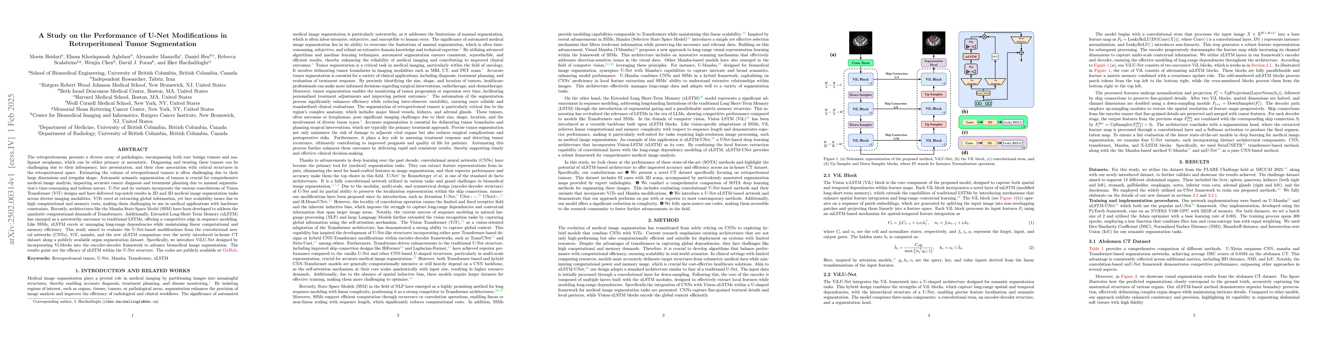
The retroperitoneum hosts a variety of tumors, including rare benign and malignant types, which pose diagnostic and treatment challenges due to their infrequency and proximity to vital structures. Estimating tumor volume is difficult due to their irregular shapes, and manual segmentation is time-consuming. Automatic segmentation using U-Net and its variants, incorporating Vision Transformer (ViT) elements, has shown promising results but struggles with high computational demands. To address this, architectures like the Mamba State Space Model (SSM) and Extended Long-Short Term Memory (xLSTM) offer efficient solutions by handling long-range dependencies with lower resource consumption. This study evaluates U-Net enhancements, including CNN, ViT, Mamba, and xLSTM, on a new in-house CT dataset and a public organ segmentation dataset. The proposed ViLU-Net model integrates Vi-blocks for improved segmentation. Results highlight xLSTM's efficiency in the U-Net framework. The code is publicly accessible on GitHub.

AI Key Findings

Generated Jun 12, 2025

Methodology

This study evaluates U-Net enhancements, including CNN, ViT, Mamba, and xLSTM, on a new in-house CT dataset and a public organ segmentation dataset. The proposed ViLU-Net model integrates Vi-blocks for improved segmentation.

Key Results

  • xLSTM's efficiency in the U-Net framework is highlighted.
  • ViLU-Net outperforms other methods, achieving average DSC scores of 0.8594 on the abdomen CT dataset.
  • ViLU-Net demonstrates superior performance on the retroperitoneal tumor dataset, with the highest average DSC, NSD, IoU, and the lowest average HD.

Significance

This research introduces a novel CT dataset for retroperitoneal tumors and evaluates state-of-the-art segmentation methods, showcasing the potential of ViLU-Net for clinical applications in precise tumor segmentation.

Technical Contribution

The ViLU-Net model, which integrates Vi-blocks into a U-shaped architecture for precise semantic segmentation, is a key technical contribution.

Novelty

This work introduces a novel ViLU-Net model that combines the strengths of ViL blocks and U-Net architecture, offering superior performance in tumor segmentation with reduced complexity compared to existing methods.

Limitations

  • The study did not explore the integration of multiple imaging modalities for a more comprehensive representation of a patient's condition.
  • The research did not address the optimization of model efficiency specifically for clinical deployment.

Future Work

  • Develop robust multi-modal fusion techniques to effectively combine information from different medical imaging sources.
  • Optimize model efficiency for clinical deployment and enhance interpretability.

Paper Details

PDF Preview

Download PDF

Similar Papers

Found 4 papers

No citations found for this paper.

Comments (0)

This website uses cookies to ensure you get the best experience. Learn more