This paper reviews recent methods for incorporating anatomical constraints into medical image segmentation, aiming to improve consistency and accuracy in downstream tasks such as surgical planning and treatment planning.
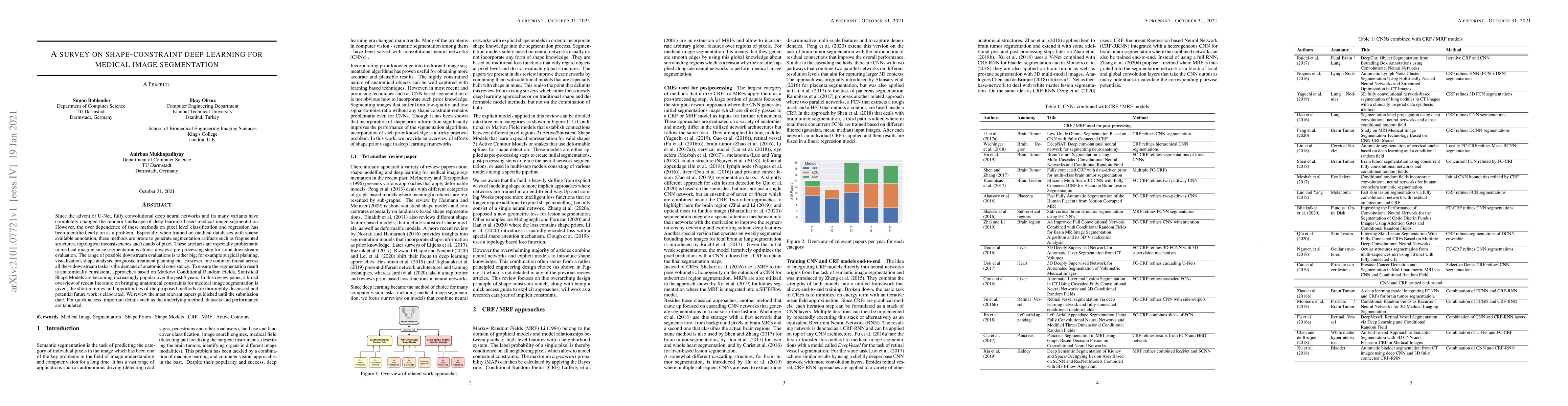
Since the advent of U-Net, fully convolutional deep neural networks and its many variants have completely changed the modern landscape of deep learning based medical image segmentation. However, the over dependence of these methods on pixel level classification and regression has been identified early on as a problem. Especially when trained on medical databases with sparse available annotation, these methods are prone to generate segmentation artifacts such as fragmented structures, topological inconsistencies and islands of pixel. These artefacts are especially problematic in medical imaging since segmentation is almost always a pre-processing step for some downstream evaluation. The range of possible downstream evaluations is rather big, for example surgical planning, visualization, shape analysis, prognosis, treatment planning etc. However, one common thread across all these downstream tasks is the demand of anatomical consistency. To ensure the segmentation result is anatomically consistent, approaches based on Markov/ Conditional Random Fields, Statistical Shape Models are becoming increasingly popular over the past 5 years. In this review paper, a broad overview of recent literature on bringing anatomical constraints for medical image segmentation is given, the shortcomings and opportunities of the proposed methods are thoroughly discussed and potential future work is elaborated. We review the most relevant papers published until the submission date. For quick access, important details such as the underlying method, datasets and performance are tabulated.
Get AI-generated insights about this paper's methodology, results, and significance.
Current paper (gray), citations (green), references (blue)
Yichi Zhang, Jicong Zhang, Rushi Jiao et al.
Jun Liu, Shengzhe Chen, Zhaoxuan Dong
Title | Authors | Year | Actions |
---|---|---|---|
Development and Analysis of an AI-Driven System for Automated Liver Segmentation in Medical Imaging using Deep Learning
|
Shruti Chauhan, Harshit Mishra, Krish Gupta, Dr Ansh Gupta, Ram Paul, S. Tomar
|
2025
|
View Paper |
World of Forms: Deformable geometric templates for one-shot surface meshing in coronary CT angiography
|
R. V. Herten, Ioannis Lagogiannis, J. Wolterink, Steffen Bruns, E. Meulendijks, Damini Dey, J. D. Groot, José P. Henriques, Nils Planken, Simone Saitta, I. Išgum
|
2025
|
View Paper |
Structural uncertainty estimation for medical image segmentation
|
Bing Yang, Xiaoqing Zhang, Huihong Zhang, Sanqian Li, Risa Higashita, Jiang Liu
|
2025
|
View Paper |
Deep generative models for vessel segmentation in CT angiography of the brain
|
H. V. Voorst, Jiahang Su, P. Konduri, C. B. Majoie, Y. Roos, B. Emmer, Henk A. Marquering, Bob D. de Vos, M. Caan, Ivana Iˇsgum, W. A. Caan
|
2025
|
View Paper |
Combining artificial intelligence assisted image segmentation and ultrasound based radiomics for the prediction of carotid plaque stability
|
Jiajia Song, Liwen Zou, Yu Li, Xiaoyin Wang, Junlan Qiu, Kailin Gong
|
2025
|
View Paper |
Multi-View and Multi-Type Feature Fusion Rotor Biofouling Recognition Method for Tidal Stream Turbine
|
Haoran Xu, Dingding Yang, Tian-zheng Wang, M. Benbouzid
|
2025
|
View Paper |
TreeFormer: Single-View Plant Skeleton Estimation via Tree-Constrained Graph Generation
|
Xinpeng Liu, Hiroaki Santo, Yosuke Toda, Fumio Okura
|
2024
|
View Paper |
A max value approach of constructing 2-D histogram for auto image multilevel segmentation
|
Yunfei Zhang, Zhan Zhang, Ri Xu, Ping Wang, Xianghong He
|
2024
|
View Paper |
Auto thresholds of image multilevel segmentation applying maximum value to construct 2D histogram
|
Yunfei Zhang, Zhan Zhang, Ri Xu, Ping Wang, Xianghong He
|
2024
|
View Paper |
World of Forms: Deformable Geometric Templates for One-Shot Surface Meshing in Coronary CT Angiography
|
R. V. Herten, Ioannis Lagogiannis, J. Wolterink, Steffen Bruns, E. Meulendijks, Damini Dey, J. D. Groot, José P. Henriques, R. N. Planken, Simone Saitta, Ivana Ivsgum
|
2024
|
View Paper |
SPGNet: A Shape-prior Guided Network for Medical Image Segmentation
|
Zhengxuan Song, Xun Liu, Wenhao Zhang, Yongyi Gong, T. Hao, Kun Zeng
|
2024
|
View Paper |
A Minimum Means of Creating 2 D Entropy for Image Multi-Value Segmentation
|
Yunfei Zhang, Zhan Zhang, Xianghong He
|
2024
|
View Paper |
Refining Biologically Inconsistent Segmentation Masks with Masked Autoencoders
|
Alexander Sauer, Yuan Tian, Joerg Bewersdorf, Jens Rittscher
|
2024
|
View Paper |
Automated Ultrasound Object Segmentation Using Combinatorial Active Contour Method
|
A. Nugroho, Budi Sunarko, Hari Wibawanto, Anggraini Mulwinda, Anas Fauzi, Dwi Oktaviyanti, Dina Wulung Savitri
|
2024
|
View Paper |
Unfolding Explainable AI for Brain Tumor Segmentation
|
Muhammad Hassan, A. A. Fateh, Jieqiong Lin, Yijiang Zhuang, Guisen Lin, Hairui Xiong, You Zhou, Peiwu Qin, Hongwu Zeng
|
2024
|
View Paper |
MCE: Medical Cognition Embedded in 3D MRI feature extraction for advancing glioma staging
|
Han Xue, Huimin Lu, Yi-ling Wang, Niya Li, Guizeng Wang
|
2024
|
View Paper |
Saliency and boundary guided segmentation framework for cell counting in microscopy images
|
S. Asha, G. Gopakumar, G. R. S. Subrahmanyam
|
2024
|
View Paper |
Benchmarking bias: Expanding clinical AI model card to incorporate bias reporting of social and non-social factors
|
C. A. M. Heming, Mohamed Abdalla, Monish Ahluwalia, Linglin Zhang, H. Trivedi, MinJae Woo, Benjamin A. Fine, J. Gichoya, L. Celi, Laleh Seyyed-Kalantari
|
2023
|
View Paper |
Enhancing Fetal Anomaly Detection in Ultrasonography Images: A Review of Machine Learning-Based Approaches
|
Ramin Yousefpour Shahrivar, Fatemeh Karami, Ebrahim Karami
|
2023
|
View Paper |
An Anatomy- and Topology-Preserving Framework for Coronary Artery Segmentation
|
Xiao Zhang, Kaicong Sun, Dijia Wu, Xiaosong Xiong, Jiameng Liu, Linlin Yao, Shufang Li, Yining Wang, Jun Feng, Dinggang Shen
|
2023
|
View Paper |
Deep neural network pulmonary nodule segmentation methods for CT images: Literature review and experimental comparisons
|
Lijia Zhi, Wujun Jiang, Shaomin Zhang, Tao Zhou
|
2023
|
View Paper |
Attractive deep morphology-aware active contour network for vertebral body contour extraction with extensions to heterogeneous and semi-supervised scenarios
|
Shen Zhao, Jinhong Wang, Xinxin Wang, Yikang Wang, Hanying Zheng, B. Chen, Antonia Zeng, Fuxin Wei, Sadeer G. Al-Kindi, S. Li
|
2023
|
View Paper |
Medical Image Segmentation Using Deep Learning: Review
|
Aida Hussein, W. Abd-Elhafiez, E. Zanaty, Mohamed Hussein
|
2023
|
View Paper |
Validated respiratory drug deposition predictions from 2D and 3D medical images with statistical shape models and convolutional neural networks
|
Josh Williams, Haavard Ahlqvist, Alexander Cunningham, Andrew Kirby, I. Katz, J. Fleming, J. Conway, Steve Cunningham, A. Ozel, U. Wolfram
|
2023
|
View Paper |
Source-Free Unsupervised Domain Adaptation with Norm and Shape Constraints for Medical Image Segmentation
|
Satoshi Kondo
|
2022
|
View Paper |
Enforcing Geometrical Priors in Deep Networks for Semantic Segmentation Applied to Radiotherapy Planning
|
Zoé Lambert, Carole Le Guyader, C. Petitjean
|
2022
|
View Paper |
Improving Anatomical Plausibility in Medical Image Segmentation via Hybrid Graph Neural Networks: Applications to Chest X-Ray Analysis
|
Nicolás Gaggion, Lucas Mansilla, Candelaria Mosquera, Diego H. Milone, Enzo Ferrante
|
2022
|
View Paper |
A novel shape-based loss function for machine learning-based seminal organ segmentation in medical imaging
|
Reza Karimzadeh, E. Fatemizadeh, H. Arabi
|
2022
|
View Paper |
A Review on Deep-Learning Algorithms for Fetal Ultrasound-Image Analysis
|
M. C. Fiorentino, Francesca Pia Villani, M. D. Cosmo, E. Frontoni, S. Moccia
|
2022
|
View Paper |
Anatomy-aided deep learning for medical image segmentation: a review
|
Lu Liu, J. Wolterink, Christoph Brune, R. Veldhuis
|
2021
|
View Paper |
TDA SegUNet: Topological Data Analysis-Based Shape-Aware Brain Tumor Segmentation
|
Ansar Rahman, Ayesha Satti, Ahmad Raza Shahid, Qaisar M. Shafi, Khadija Farooq, Asad Ali Safi
|
2025
|
View Paper |
A Surprisingly Effective Perimeter-based Loss for Medical Image Segmentation
|
Rosana El Jurdi, C. Petitjean, V. Cheplygina, F. Abdallah
|
2021
|
View Paper |
Shape Constraints in Deep Learning for Robust 2D Echocardiography Analysis
|
Yingyu Yang, Maxime Sermesant
|
2021
|
View Paper |
Computerized Medical Imaging and Graphics
|
A. Sadikine, Bogdan Badic, J.-P. Tasu, Vincent Noblet, P. Ballet, Dimitris Visvikis, Pierre-Henri Conze
|
View Paper |
Comments (0)