Publication
Metrics
AI Quick Summary
A new contrast mechanism for scanning transmission electron microscopy enhances atomic resolution imaging by detecting symmetry in specimen exit electron intensity distributions, revealing sharp peaks for atomic columns with improved sensitivity and precision.
Paper Preview
Abstract
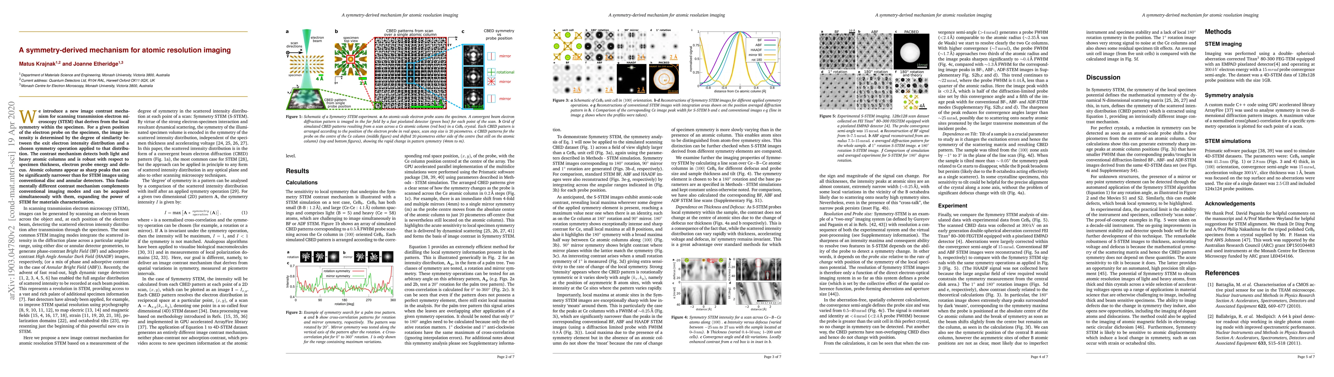
We introduce a new image contrast mechanism for scanning transmission electron microscopy (STEM) that derives from the local symmetry within the specimen. For a given position of the electron probe on the specimen, the image intensity is determined by the degree of similarity between the exit electron intensity distribution and a chosen symmetry operation applied to that distribution. The contrast mechanism detects both light and heavy atomic columns and is robust with respect to specimen thickness, electron probe energy and defocus. Atomic columns appear as sharp peaks that can be significantly narrower than for STEM images using conventional disc and annular detectors. This fundamentally different contrast mechanism complements conventional imaging modes and can be acquired simultaneously with them, expanding the power of STEM for materials characterisation.
AI Key Findings
Get AI-generated insights about this paper's methodology, results, significance, and more — seven facets brought into focus.
Impact
Paper Details
Authors
PDF Preview
Key Terms
Citation Network
Current paper (gray), citations (green), references (blue)
Display is limited for performance on very large graphs.
Discussion 0