A Systematic Approach for MRI Brain Tumor Localization, and Segmentation using Deep Learning and Active Contouring
Publication
Metrics
Paper Preview
Abstract
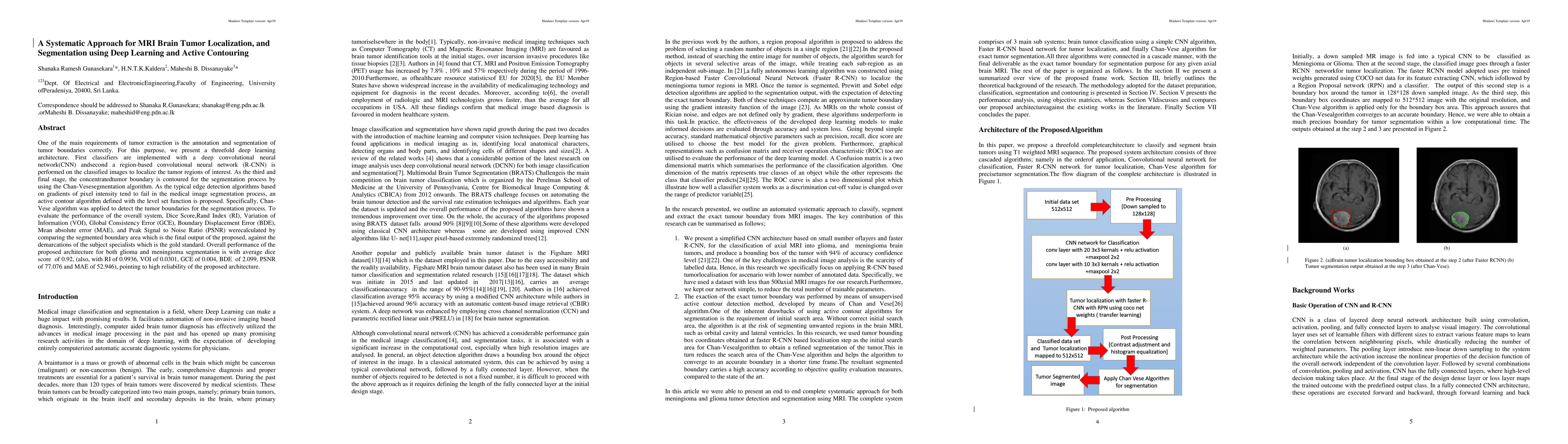
One of the main requirements of tumor extraction is the annotation and segmentation of tumor boundaries correctly. For this purpose, we present a threefold deep learning architecture. First classifiers are implemented with a deep convolutional neural network(CNN) andsecond a region-based convolutional neural network (R-CNN) is performed on the classified images to localize the tumor regions of interest. As the third and final stage, the concentratedtumor boundary is contoured for the segmentation process by using the Chan-Vesesegmentation algorithm. As the typical edge detection algorithms based on gradients of pixel intensity tend to fail in the medical image segmentation process, an active contour algorithm defined with the level set function is proposed. Specifically, Chan- Vese algorithm was applied to detect the tumor boundaries for the segmentation process. To evaluate the performance of the overall system, Dice Score,Rand Index (RI), Variation of Information (VOI), Global Consistency Error (GCE), Boundary Displacement Error (BDE), Mean absolute error (MAE), and Peak Signal to Noise Ratio (PSNR) werecalculated by comparing the segmented boundary area which is the final output of the proposed, against the demarcations of the subject specialists which is the gold standard. Overall performance of the proposed architecture for both glioma and meningioma segmentation is with average dice score of 0.92, (also, with RI of 0.9936, VOI of 0.0301, GCE of 0.004, BDE of 2.099, PSNR of 77.076 and MAE of 52.946), pointing to high reliability of the proposed architecture.
AI Key Findings
Get AI-generated insights about this paper's methodology, results, significance, and more — seven facets brought into focus.
Impact
Paper Details
Authors
PDF Preview
Key Terms
Citation Network
Current paper (gray), citations (green), references (blue)
Display is limited for performance on very large graphs.
Discussion 0