This research develops a semi-supervised learning framework for medical image segmentation using a teacher-student approach with partial dense labels and easier bounding-box annotations. It introduces an organ-to-lesion attention module and localization branch to enhance segmentation performance, achieving state-of-the-art results on LiTS challenge datasets.
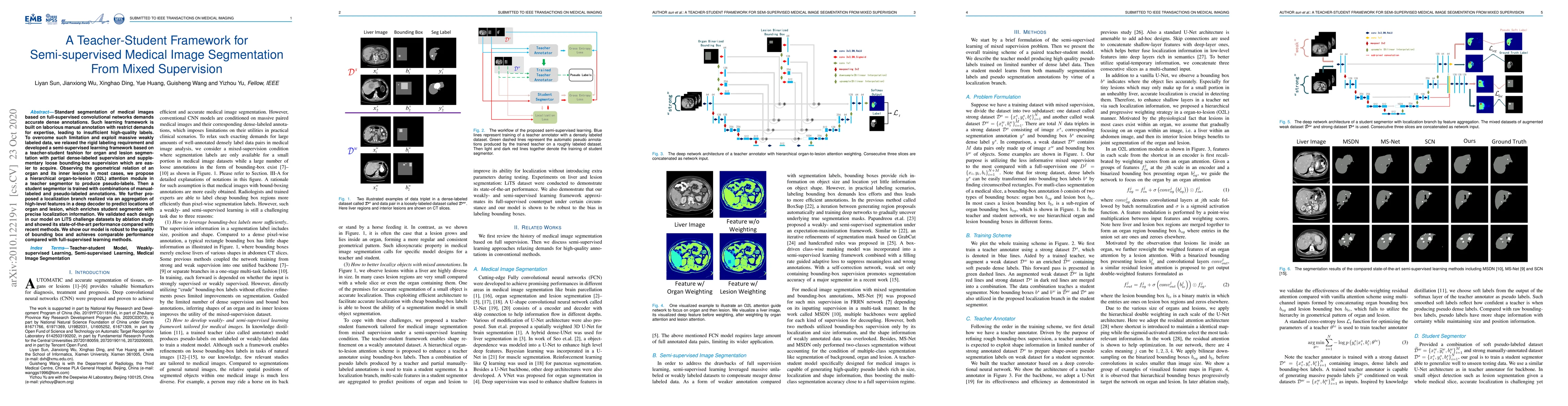
Standard segmentation of medical images based on full-supervised convolutional networks demands accurate dense annotations. Such learning framework is built on laborious manual annotation with restrict demands for expertise, leading to insufficient high-quality labels. To overcome such limitation and exploit massive weakly labeled data, we relaxed the rigid labeling requirement and developed a semi-supervised learning framework based on a teacher-student fashion for organ and lesion segmentation with partial dense-labeled supervision and supplementary loose bounding-box supervision which are easier to acquire. Observing the geometrical relation of an organ and its inner lesions in most cases, we propose a hierarchical organ-to-lesion (O2L) attention module in a teacher segmentor to produce pseudo-labels. Then a student segmentor is trained with combinations of manual-labeled and pseudo-labeled annotations. We further proposed a localization branch realized via an aggregation of high-level features in a deep decoder to predict locations of organ and lesion, which enriches student segmentor with precise localization information. We validated each design in our model on LiTS challenge datasets by ablation study and showed its state-of-the-art performance compared with recent methods. We show our model is robust to the quality of bounding box and achieves comparable performance compared with full-supervised learning methods.
This graph shows how this paper is connected to other research. The current paper is shown in dark gray, papers that cite it are in green, and papers it references are in blue. You can:
• Hover over any node to see the paper title
• Click a node to view that paper's details
• Drag nodes to rearrange the graph
• Use the zoom controls (bottom right) to zoom in/out
Title | Authors | Year | Actions |
---|---|---|---|
Identifying retinopathy in optical coherence tomography images with less labeled data via contrastive graph regularization
|
Songqi Hu, Hongying Tang, Yuemei Luo
|
2024
|
View Paper |
Beyond Pixel-Wise Supervision for Medical Image Segmentation: From Traditional Models to Foundation Models
|
Yuyan Shi, Jialu Ma, Jin Yang, Shasha Wang, Yichi Zhang
|
2024
|
View Paper |
Sd-net: a semi-supervised double-cooperative network for liver segmentation from computed tomography (CT) images
|
Shixin Huang, Jiawei Luo, Yangning Ou, Wangjun shen, Yu Pang, Xixi Nie, Guo Zhang
|
2024
|
View Paper |
Sensitivity-guided iterative parameter identification and data generation with BayesFlow and PELS-VAE for model calibration
|
Yi Zhang, Lars Mikelsons
|
2023
|
View Paper |
Weakly-Interactive-Mixed Learning: Less Labelling Cost for Better Medical Image Segmentation
|
Xiuping Nie, Lilu Liu, Lifeng He, Liang Zhao, Haojian Lu, Songmei Lou, R. Xiong, Yue Wang
|
2023
|
View Paper |
Fast, light, and scalable: harnessing data-mined line annotations for automated tumor segmentation on brain MRI
|
Nathaniel C Swinburne, V. Yadav, K. Murthy, Pierre Elnajjar, Hao-Hsin Shih, P. Panyam, A. Santilli, David C Gutman, L. Pike, N. Moss, J. Stone, V. Hatzoglou, A. Shah, K. Juluru, Sohrab P. Shah, A. Holodny, R. Young, For The M.S.K. Mind Consortium
|
2023
|
View Paper |
Self-training with dual uncertainty for semi-supervised medical image segmentation
|
Zhanhong Qiu, Haitao Gan, Mingzhi Shi, Zhongwei Huang, Zhi Yang
|
2023
|
View Paper |
A Radiogenomics Pipeline for Lung Nodules Segmentation and Prediction of EGFR Mutation Status from CT Scans
|
Ivo Gollini Navarrete, Mohammad Yaqub
|
2022
|
View Paper |
Efficient Combination of CNN and Transformer for Dual-Teacher Uncertainty-guided Semi-supervised Medical Image Segmentation
|
Zhiyong Xiao, Yixin Su, Zhaohong Deng, W. Zhang
|
2022
|
View Paper |
SCANet: A Unified Semi-Supervised Learning Framework for Vessel Segmentation
|
Ning Shen, Tingfa Xu, Ziyang Bian, Shiqi Huang, Feng Mu, Bo Huang, Yuze Xiao, Jianan Li
|
2022
|
View Paper |
CAFS: An Attention-Based Co-Segmentation Semi-Supervised Method for Nasopharyngeal Carcinoma Segmentation
|
Yitong Chen, Guanghui Han, Tianyu Lin, Xiujian Liu
|
2022
|
View Paper |
Teacher-student approach for lung tumor segmentation from mixed-supervised datasets
|
Vemund Fredriksen, S. O. M. Sevle, A. Pedersen, T. Langø, G. Kiss, F. Lindseth
|
2022
|
View Paper |
Teacher-Student Architecture for Mixed Supervised Lung Tumor Segmentation
|
Vemund Fredriksen, Svein Ole M. Svele, A. Pedersen, T. Langø, G. Kiss, F. Lindseth
|
2021
|
View Paper |
Semi-supervised 3D Object Detection via Temporal Graph Neural Networks
|
Jianren Wang, Haiming Gang, Siddarth Ancha, Yi-Ting Chen, David Held
|
2021
|
View Paper |
Inconsistency-Aware Uncertainty Estimation for Semi-Supervised Medical Image Segmentation
|
Yinghuan Shi, Jian Zhang, T. Ling, Jiwen Lu, Yefeng Zheng, Qian Yu, Lei Qi, Yang Gao
|
2021
|
View Paper |
Yichi Zhang, Yuyan Shi, Shasha Wang
Published: 2023-10-24
Published: 2020-08-10
Zhen Zhao, Yixuan Yuan, Dian Yu, Longyue Wang, Zicheng Wang, Luping Zhou
Published: 2023-11-29
Gorkem Durak, Ulas Bagci, Yuri S. Velichko, Koushik Biswas, Meghana Karri, Vedat Cicek, Amit Soni Arya, Nicol`o Gennaro
Published: 2024-12-19
Comments (0)