01
MethodologyHow they did it
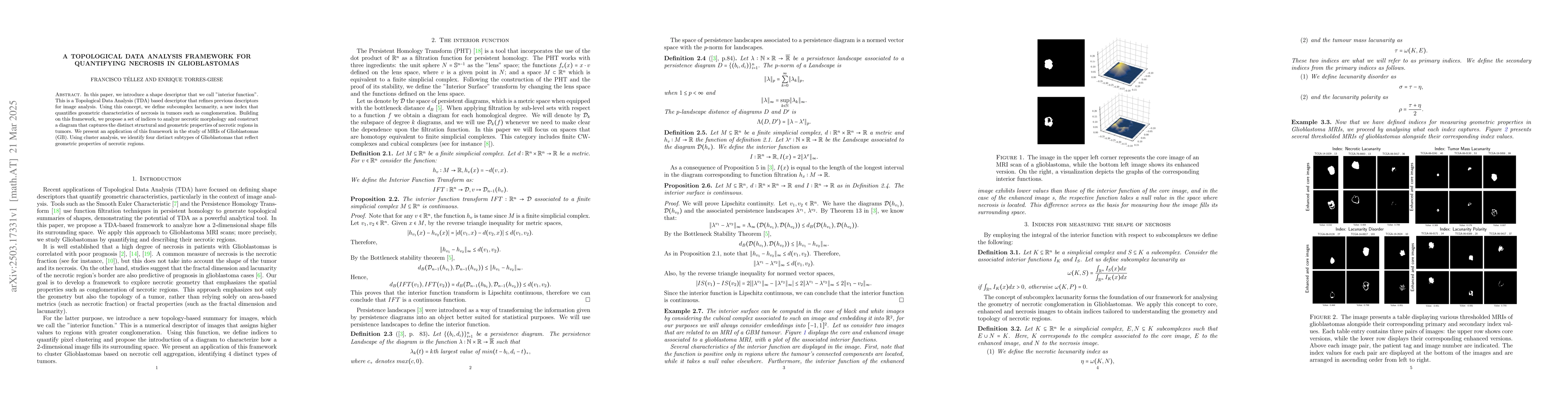
The research introduces a Topological Data Analysis (TDA) based shape descriptor called 'interior function' to quantify necrosis in tumors. It defines subcomplex lacunarity, a new index, and constructs a primary index diagram to capture distinct structural and geometric properties of necrotic regions in tumors.
Discussion 0