A unified image reconstruction framework for quantitative dual- and triple-energy CT imaging of material compositions
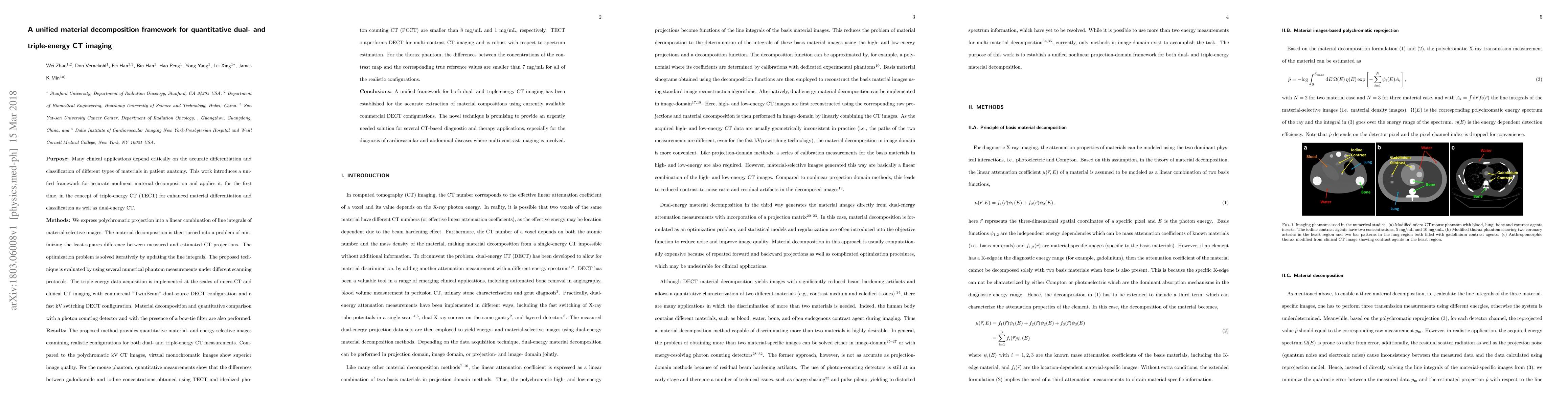
Publication
Metrics
AI Quick Summary
This paper presents a unified framework for accurate material decomposition in dual- and triple-energy CT imaging, demonstrating superior material differentiation and classification compared to traditional methods. The proposed technique shows promising results for clinical applications, providing enhanced image quality and precise material concentration measurements.
Paper Preview
Abstract
Many clinical applications depend critically on the accurate differentiation and classification of different types of materials in patient anatomy. This work introduces a unified framework for accurate nonlinear material decomposition and applies it, for the first time, in the concept of triple-energy CT (TECT) for enhanced material differentiation and classification as well as dual-energy CT. The triple-energy data acquisition is implemented at the scales of micro-CT and clinical CT imaging with commercial "TwinBeam" dual-source DECT configuration and a fast kV switching DECT configuration. Material decomposition and quantitative comparison with a photon counting detector and with the presence of a bow-tie filter are also performed. The proposed method provides quantitative material- and energy-selective images examining realistic configurations for both dual- and triple-energy CT measurements. Compared to the polychromatic kV CT images, virtual monochromatic images show superior image quality. For the mouse phantom, quantitative measurements show that the differences between gadodiamide and iodine concentrations obtained using TECT and idealized photon counting CT (PCCT) are smaller than 8 mg/mL and 1 mg/mL, respectively. TECT outperforms DECT for multi-contrast CT imaging and is robust with respect to spectrum estimation. For the thorax phantom, the differences between the concentrations of the contrast map and the corresponding true reference values are smaller than 7 mg/mL for all of the realistic configurations. A unified framework for both dual- and triple-energy CT imaging has been established for the accurate extraction of material compositions; considering currently available commercial DECT configurations. The novel technique is promising to provide an urgently needed solution for several CT-based diagnosis and therapy applications.
AI Key Findings
Get AI-generated insights about this paper's methodology, results, significance, and more — seven facets brought into focus.
Impact
Paper Details
PDF Preview
Key Terms
Citation Network
Current paper (gray), citations (green), references (blue)
Display is limited for performance on very large graphs.
Discussion 0