A Unified Multi-Scale Attention-Based Network for Automatic 3D Segmentation of Lung Parenchyma & Nodules In Thoracic CT Images

Source: ArXiv

AI Quick Summary

A new attention-based network for 3D segmentation of lung parenchyma and nodules in thoracic CT images achieves better performance than state-of-the-art methods with improved accuracy and robustness.

Share: Share on X LinkedIn

Summary

Lung cancer has been one of the major threats across the world with the highest mortalities. Computer-aided detection (CAD) can help in early detection and thus can help increase the survival rate. Accurate lung parenchyma segmentation (to include the juxta-pleural nodules) and lung nodule segmentation, the primary symptom of lung cancer, play a crucial role in the overall accuracy of the Lung CAD pipeline. Lung nodule segmentation is quite challenging because of the diverse nodule types and other inhibit structures present within the lung lobes. Traditional machine/deep learning methods suffer from generalization and robustness. Recent Vision Language Models/Foundation Models perform well on the anatomical level, but they suffer on fine-grained segmentation tasks, and their semi-automatic nature limits their effectiveness in real-time clinical scenarios. In this paper, we propose a novel method for 
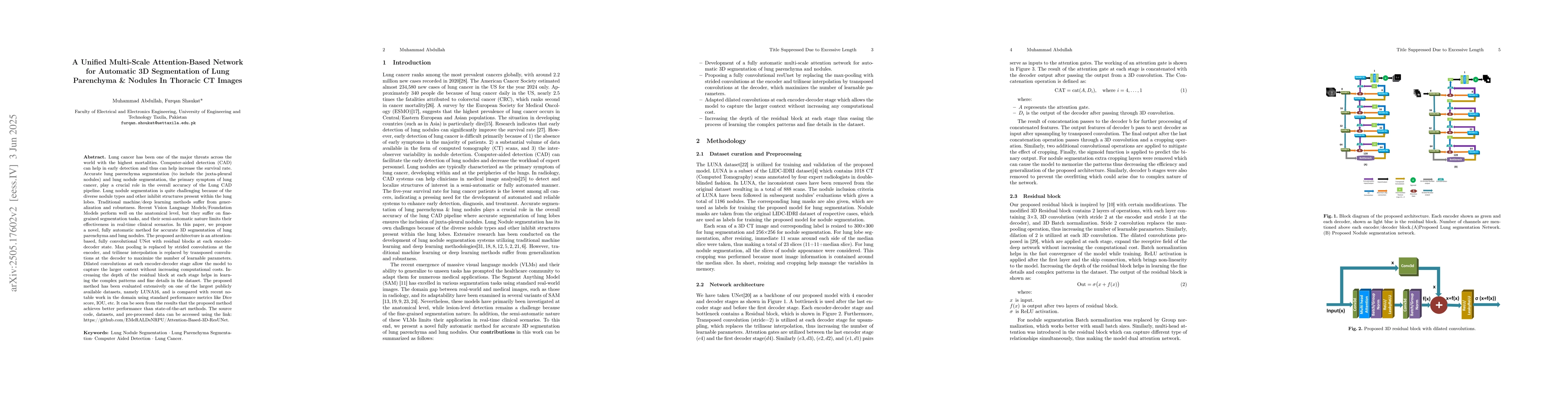
accurate 3D segmentation of lung parenchyma and lung nodules. The proposed architecture is an attention-based network with residual blocks at each encoder-decoder state. Max pooling is replaced by strided convolutions at the encoder, and trilinear interpolation is replaced by transposed convolutions at the decoder to maximize the number of learnable parameters. Dilated convolutions at each encoder-decoder stage allow the model to capture the larger context without increasing computational costs. The proposed method has been evaluated extensively on one of the largest publicly available datasets, namely LUNA16, and is compared with recent notable work in the domain using standard performance metrics like Dice score, IOU, etc. It can be seen from the results that the proposed method achieves better performance than state-of-the-art methods. The source code, datasets, and pre-processed data can be accessed using the link: https://github.com/EMeRALDsNRPU/Attention-Based-3D-ResUNet.

AI Key Findings

Generated Jun 07, 2025

Methodology

The research proposes a novel attention-based network with residual blocks at each encoder-decoder state. It replaces max pooling with strided convolutions at the encoder and trilinear interpolation with transposed convolutions at the decoder. Dilated convolutions are employed to capture larger context without increasing computational costs.

Key Results

  • The proposed method outperforms state-of-the-art techniques in 3D segmentation of lung parenchyma and nodules as per extensive evaluation on the LUNA16 dataset.
  • Standard performance metrics such as Dice score and IOU demonstrate superior results achieved by the proposed architecture.

Significance

This research is crucial for improving early lung cancer detection by providing accurate 3D segmentation of lung parenchyma and nodules, which is essential for computer-aided detection (CAD) systems.

Technical Contribution

The main technical contribution is a unified multi-scale attention-based network architecture with residual connections, dilated convolutions, and specific replacements of pooling/interpolation layers for efficient 3D segmentation.

Novelty

This work stands out by combining multi-scale attention mechanisms with residual blocks in a 3D U-Net architecture, specifically tailored for lung parenchyma and nodule segmentation, addressing challenges posed by diverse nodule types and complex lung structures.

Limitations

  • The paper does not discuss limitations explicitly, but potential limitations could include reliance on large datasets for training and computational resources for the complex network architecture.

Future Work

  • Exploration of methods to further reduce computational costs while maintaining or improving accuracy.
  • Investigating the applicability of the proposed method on other medical imaging modalities beyond thoracic CT scans.

Paper Details

PDF Preview

Download PDF

Citation Network

Current paper (gray), citations (green), references (blue)

Display is limited for performance on very large graphs.

Current Paper
Citations
References
Click to view

No citations found for this paper.

Comments (0)

This website uses cookies to ensure you get the best experience. Learn more