Publication
Metrics
AI Quick Summary
This paper introduces a whole-slide image grading benchmark for cervical cancer precursor lesions, addressing papillae and cell overlap issues, and develops a tissue classification method using novel morphological features. It also evaluates inter-observer variability among pathologists in diagnosing these lesions.
Paper Preview
Abstract
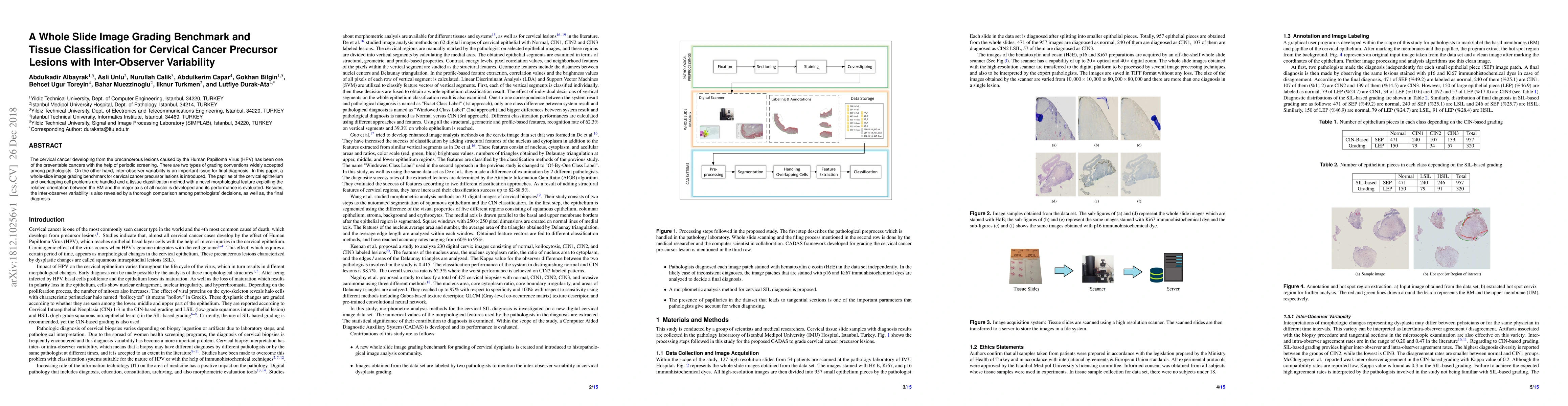
The cervical cancer developing from the precancerous lesions caused by the Human Papilloma Virus (HPV) has been one of the preventable cancers with the help of periodic screening. There are two types of grading conventions widely accepted among pathologists. On the other hand, inter-observer variability is an important issue for final diagnosis. In this paper, a whole-slide image grading benchmark for cervical cancer precursor lesions is introduced. The papillae of the cervical epithelium and overlapping cell problems are handled and a tissue classification method with a novel morphological feature exploiting the relative orientation between the BM and the major axis of all nuclei is developed and its performance is evaluated. Besides, the inter-observer variability is also revealed by a thorough comparison among pathologists' decisions, as well as, the final diagnosis.
AI Key Findings
Get AI-generated insights about this paper's methodology, results, significance, and more — seven facets brought into focus.
Impact
Paper Details
Authors
PDF Preview
Key Terms
Citation Network
Current paper (gray), citations (green), references (blue)
Display is limited for performance on very large graphs.
Discussion 0