Accelerating COVID-19 Differential Diagnosis with Explainable Ultrasound Image Analysis
Publication
Metrics
AI Quick Summary
This paper presents a deep learning approach for COVID-19 differential diagnosis using lung ultrasound images, featuring a large dataset of 106 videos. The proposed frame-based convolutional neural network achieves high sensitivity and specificity, and employs class activation maps for interpretability, validated by medical experts.
Paper Preview
Abstract
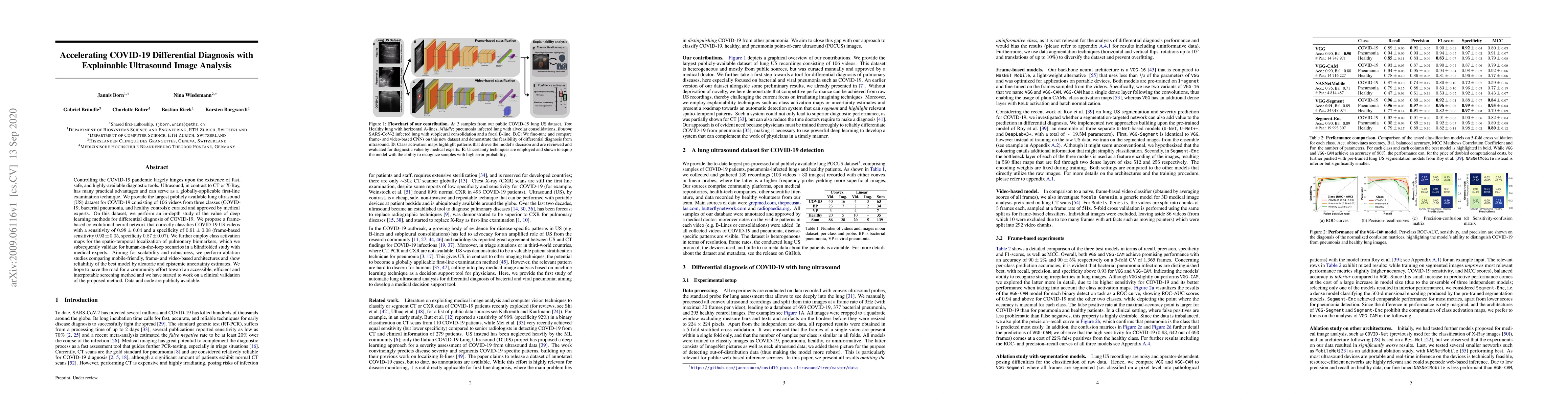
Controlling the COVID-19 pandemic largely hinges upon the existence of fast, safe, and highly-available diagnostic tools. Ultrasound, in contrast to CT or X-Ray, has many practical advantages and can serve as a globally-applicable first-line examination technique. We provide the largest publicly available lung ultrasound (US) dataset for COVID-19 consisting of 106 videos from three classes (COVID-19, bacterial pneumonia, and healthy controls); curated and approved by medical experts. On this dataset, we perform an in-depth study of the value of deep learning methods for differential diagnosis of COVID-19. We propose a frame-based convolutional neural network that correctly classifies COVID-19 US videos with a sensitivity of 0.98+-0.04 and a specificity of 0.91+-08 (frame-based sensitivity 0.93+-0.05, specificity 0.87+-0.07). We further employ class activation maps for the spatio-temporal localization of pulmonary biomarkers, which we subsequently validate for human-in-the-loop scenarios in a blindfolded study with medical experts. Aiming for scalability and robustness, we perform ablation studies comparing mobile-friendly, frame- and video-based architectures and show reliability of the best model by aleatoric and epistemic uncertainty estimates. We hope to pave the road for a community effort toward an accessible, efficient and interpretable screening method and we have started to work on a clinical validation of the proposed method. Data and code are publicly available.
AI Key Findings
Get AI-generated insights about this paper's methodology, results, significance, and more — seven facets brought into focus.
Impact
Paper Details
Authors
PDF Preview
Key Terms
Citation Network
Current paper (gray), citations (green), references (blue)
Display is limited for performance on very large graphs.
Discussion 0