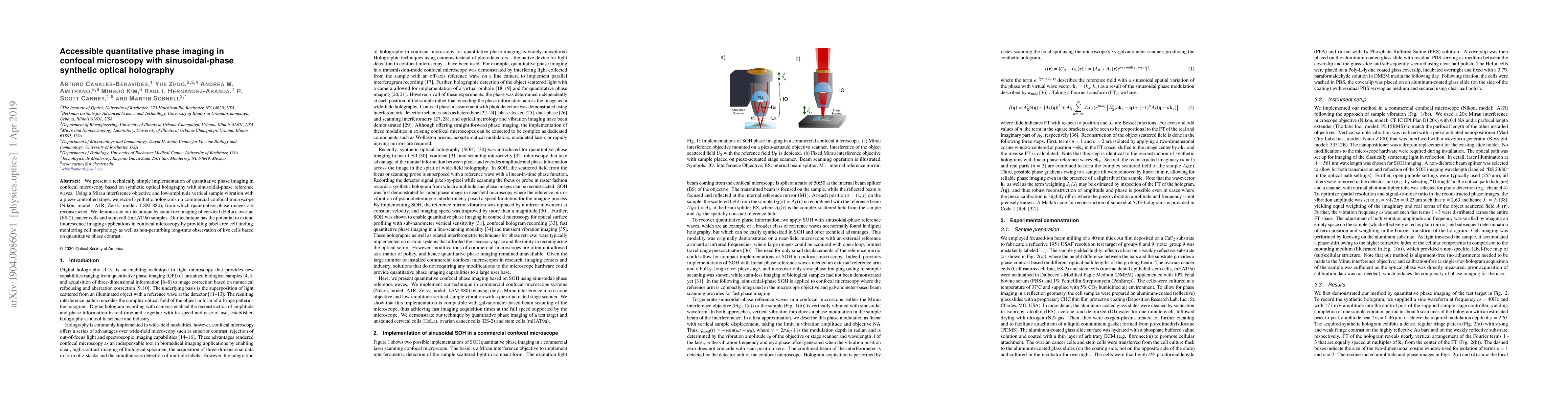
Publication
Metrics
AI Quick Summary
This paper introduces a straightforward method for quantitative phase imaging in confocal microscopy using synthetic optical holography with sinusoidal-phase reference waves. The technique enables label-free imaging of live cells, demonstrated through stain-free visualization of cervical and ovarian cancer cells, and stem cells, enhancing non-invasive cell observation.
Paper Preview
Abstract
We present a technically simple implementation of quantitative phase imaging in confocal microscopy based on synthetic optical holography with sinusoidal-phase reference waves. Using a Mirau interference objective and low-amplitude vertical sample vibration with a piezo-controlled stage, we record synthetic holograms on commercial confocal microscopes (Nikon, model: A1R; Zeiss: model: LSM-880), from which quantitative phase images are reconstructed. We demonstrate our technique by stain-free imaging of cervical (HeLa) and ovarian (ES-2) cancer cells and stem cell (mHAT9a) samples. Our technique has the potential to extend fluorescence imaging applications in confocal microscopy by providing label-free cell finding, monitoring cell morphology, as well as non-perturbing long-time observation of live cells based on quantitative phase contrast.
AI Key Findings
Get AI-generated insights about this paper's methodology, results, significance, and more — seven facets brought into focus.
Impact
Paper Details
PDF Preview
Key Terms
Citation Network
Current paper (gray), citations (green), references (blue)
Display is limited for performance on very large graphs.
Discussion 0