Active skeleton for bacteria modeling
Publication
Metrics
Paper Preview
Abstract
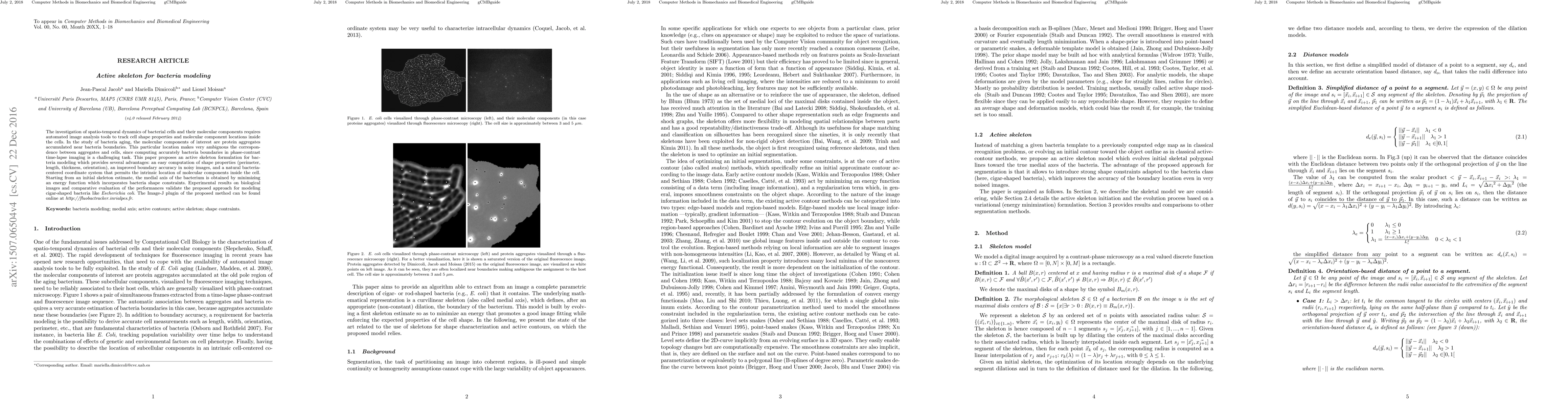
The investigation of spatio-temporal dynamics of bacterial cells and their molecular components requires automated image analysis tools to track cell shape properties and molecular component locations inside the cells. In the study of bacteria aging, the molecular components of interest are protein aggregates accumulated near bacteria boundaries. This particular location makes very ambiguous the correspondence between aggregates and cells, since computing accurately bacteria boundaries in phase-contrast time-lapse imaging is a challenging task. This paper proposes an active skeleton formulation for bacteria modeling which provides several advantages: an easy computation of shape properties (perimeter, length, thickness, orientation), an improved boundary accuracy in noisy images, and a natural bacteria-centered coordinate system that permits the intrinsic location of molecular components inside the cell. Starting from an initial skeleton estimate, the medial axis of the bacterium is obtained by minimizing an energy function which incorporates bacteria shape constraints. Experimental results on biological images and comparative evaluation of the performances validate the proposed approach for modeling cigar-shaped bacteria like Escherichia coli. The Image-J plugin of the proposed method can be found online at http://fluobactracker.inrialpes.fr.
AI Key Findings
Get AI-generated insights about this paper's methodology, results, significance, and more — seven facets brought into focus.
Impact
Paper Details
PDF Preview
Key Terms
Citation Network
Current paper (gray), citations (green), references (blue)
Display is limited for performance on very large graphs.
Discussion 0