ArxivLens

Quick Summary:

Adorym is a versatile x-ray image reconstruction framework utilizing automatic differentiation for optimization, supporting various imaging methods and parallel processing. It refines experimental parameters and demonstrates improved image quality across multiple datasets.

DOI: 10.1364/OE.418296
arXiv Id: 2012.12686
Date Published: 2021-04-07
Date Updated: 2021-04-07

Key Terms (TF-IDF):

allowing (0.224)
allowing (0.222)
optimization (0.196)
framework (0.194)
optimization (0.193)
image (0.192)
framework (0.190)
datasets (0.186)
image (0.186)
datasets (0.184)
scan (0.182)
differentiation (0.180)
differentiation (0.179)
high performance (0.177)
fly (0.174)
processed (0.172)
refine (0.172)
refinement (0.172)
scan (0.172)
high performance (0.172)
License: http://creativecommons.org/licenses/by/4.0/

Summary:

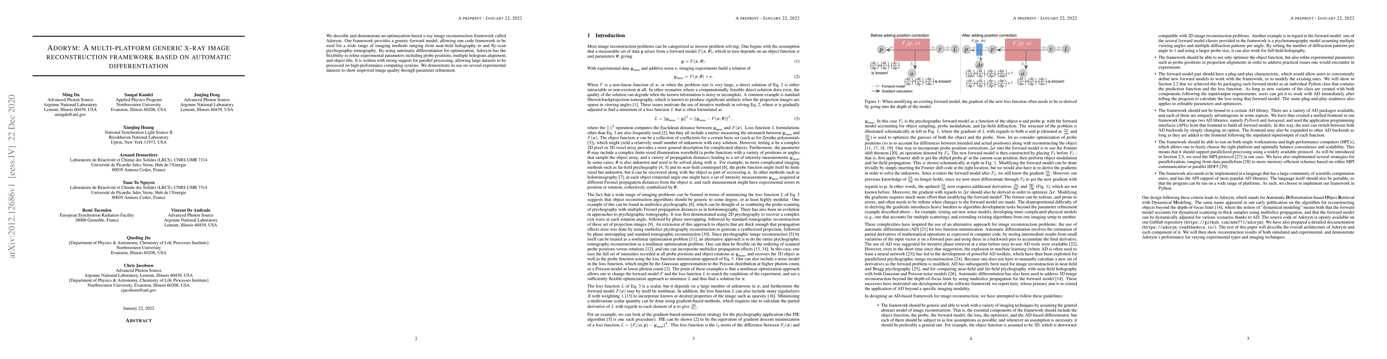
We describe and demonstrate an optimization-based x-ray image reconstruction framework called Adorym. Our framework provides a generic forward model, allowing one code framework to be used for a wide range of imaging methods ranging from near-field holography to and fly-scan ptychographic tomography. By using automatic differentiation for optimization, Adorym has the flexibility to refine experimental parameters including probe positions, multiple hologram alignment, and object tilts. It is written with strong support for parallel processing, allowing large datasets to be processed on high-performance computing systems. We demonstrate its use on several experimental datasets to show improved image quality through parameter refinement.

Citation & Reference Network Visualization

This graph shows how this paper is connected to other research. The current paper is shown in dark gray, papers that cite it are in green, and papers it references are in blue. You can:

Current Paper
Papers citing this work (Citations)
Papers cited by this work (References)

• Hover over any node to see the paper title

• Click a node to view that paper's details

• Drag nodes to rearrange the graph

• Use the zoom controls (bottom right) to zoom in/out

Citations and References

Title Authors Year Actions
ssc-cdi: A Memory-Efficient, Multi-GPU Package for Ptychography with Extreme Data
Y. R. Tonin, A. Z. Peixinho, Mauro Luiz Brandão Junior, Paola Ferraz, E. Miqueles
2024
View Paper
Comparison of Soft X-ray Spectro-Ptychography and Scanning Transmission X-ray Microscopy
A. Hitchcock, Chunyang Zhang, Haytham Eraky, Drew Higgins, Rachid Belkhou, Nicolas Millle, S. Swaraj, Stefan Stanescu, Tianxiao Sun, Jian Wang
2024
View Paper
Predicting ptychography probe positions using single-shot phase retrieval neural network
Ming Du, Tao Zhou, Junjing Deng, Daniel J. Ching, S. Henke, Mathew J. Cherukara
2024
View Paper
How auto-differentiation can improve CT workflows: classical algorithms in a modern framework.
R. Schoonhoven, Alexander Skorikov, Willem Palenstijn, Daniël Pelt, A. Hendriksen, joost batenburg
2024
View Paper
Imaging extended single crystal lattice distortion fields with multi-peak Bragg ptychography
S. Kandel, S. Maddali, Marc Allain, Xiaojing Huang, Y. Nashed, Chris Jacobsen, S. Hruszkewycz
2023
View Paper
Wavelength-multiplexed multi-mode EUV reflection ptychography based on automatic differentiation
Yifeng Shao, Sven Weerdenburg, Jacob Seifert, H. P. Urbach, A. Mosk, W. Coene
2023
View Paper
Reconstructing Lattice Vibrations of Crystals with Electron Ptychography
A. Gladyshev, Benedikt Haas, T. M. Boland, Peter Rez, Christoph T. Koch
2023
View Paper
Resolution of Virtual Depth Sectioning from Four-Dimensional Scanning Transmission Electron Microscopy.
E. Terzoudis-Lumsden, T. Petersen, H. Brown, P. Pelz, C. Ophus, S. Findlay
2023
View Paper
Determining the Projected Crystal Structure from Four-dimensional Scanning Transmission Electron Microscopy via the Scattering Matrix.
A. Sadri, S. Findlay
2023
View Paper
Automatic Differentiation for Inverse Problems in X-ray Imaging and Microscopy
Francesco Guzzi, A. Gianoncelli, F. Billè, Sergio Carrato, G. Kourousias
2023
View Paper
Differentiable Uncalibrated Imaging
Sidharth Gupta, K. Kothari, Valentin Debarnot, Ivan Dokmani'c
2022
View Paper
Lossy Compression of Electron Diffraction Patterns for Ptychography via Change of Basis
A. Gladyshev, T. Pekin, M. Schloz, B. Haas, Johannes Muller, C. Koch
2022
View Paper
Single-shot ptychography at a soft X-ray free-electron laser
K. Kharitonov, M. Mehrjoo, M. Ruiz-Lopez, B. Keitel, S. Kreis, S. Gang, R. Pan, A. Marras, J. Correa, C. Wunderer, E. Plönjes
2022
View Paper
A Method for High-Resolution Three-Dimensional Reconstruction with Ewald Sphere Curvature Correction from Transmission Electron Images
T. Gureyev, D. Paganin, H. Brown, H. Quiney, L. Allen
2022
View Paper
Recovering the phase and amplitude of X-ray FEL pulses using neural networks and differentiable models.
D. Ratner, F. Christie, J. Cryan, A. Edelen, A. Lutman, X. Zhang
2021
View Paper
A parameter refinement method for Ptychography based on Deep Learning concepts
Francesco Guzzi, G. Kourousias, F. Billè, R. Pugliese, A. Gianoncelli, S. Carrato
2021
View Paper
Efficient ptychographic phase retrieval via a matrix-free Levenberg-Marquardt algorithm.
S. Kandel, S. Maddali, Y. Nashed, S. Hruszkewycz, C. Jacobsen, M. Allain
2021
View Paper
Using a modified double deep image prior for crosstalk mitigation in multislice ptychography
Ming Du, Xiaojing Huang, C. Jacobsen
2021
View Paper
Gradient-based strategy of designing cascaded phase holograms for arbitrary distribution behind arbitrary aperture
Hengyang Li, Jiaming Xu, Qin Luo, Cong Hu, Yu Xiao, Xiahui Tang, Gang Xu, Yingxiong Qin
View Paper
Lensless X-Ray Nanoimaging
©SHUTTERSTOCK.COM, Tomatito
View Paper

Comments (0)

This website uses cookies to ensure you get the best experience. Learn more