Advanced 3D-Printed Multiphasic Scaffold with Optimal PRP Dosage for Chondrogenesis of BM-MSCs in Osteochondral Tissue Engineering

Source: ArXiv

AI Quick Summary

This paper develops advanced 3D-printed multiphasic scaffolds using alginate, gelatin, and varying concentrations of graphene oxide and platelet-rich plasma for osteochondral tissue engineering. Optimal PRP dosage of 2% enhances BM-MSC chondrogenesis, indicating the scaffold's potential in regenerating both bone and cartilage tissues.

Share: Share on X LinkedIn

Summary

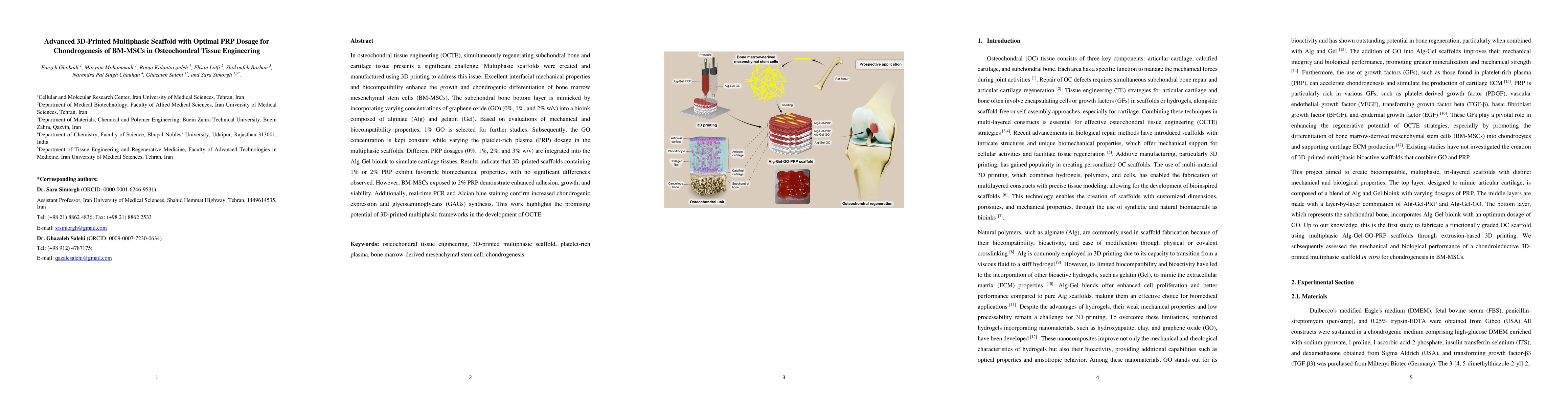
In osteochondral tissue engineering (OCTE), simultaneously regenerating subchondral bone and cartilage tissue presents a significant challenge. Multiphasic scaffolds were created and manufactured using 3D printing to address this issue. Excellent interfacial mechanical properties and biocompatibility enhance the growth and chondrogenic differentiation of bone marrow mesenchymal stem cells (BM-MSCs). The subchondral bone bottom layer is mimicked by incorporating varying concentrations of graphene oxide (GO) (0%, 1%, and 2% w/v) into a bioink composed of alginate (Alg) and gelatin (Gel). Based on evaluations of mechanical and biocompatibility properties, 1% GO is selected for further studies. Subsequently, the GO concentration is kept constant while varying the platelet-rich plasma (PRP) dosage in the multiphasic scaffolds. Different PRP dosages (0%, 1%, 2%, and 3% w/v) are integrated into the Alg-Gel bioink to simulate cartilage tissues. Results indicate that 3D-printed scaffolds containing 1% or 2% PRP exhibit favorable biomechanical properties, with no significant differences observed. However, BM-MSCs exposed to 2% PRP demonstrate enhanced adhesion, growth, and viability. Additionally, real-time PCR and Alcian blue staining confirm increased chondrogenic expression and glycosaminoglycans (GAGs) synthesis. This work highlights the promising potential of 3D-printed multiphasic frameworks in the development of OCTE.

AI Key Findings

Generated Jun 11, 2025

Methodology

The study utilized extrusion-based multi-nozzle 3D printing to create tri-layered multiphasic scaffolds mimicking osteochondral (OC) tissue, incorporating varying concentrations of graphene oxide (GO) for the subchondral bone layer and platelet-rich plasma (PRP) for the cartilage layer, followed by in vitro biocompatibility evaluation with bone marrow-derived mesenchymal stem cells (BM-MSCs).

Key Results

  • 1% GO concentration in the subchondral layer bioink provided optimal mechanical properties and biocompatibility.
  • 3D-printed scaffolds with 1% or 2% PRP exhibited favorable biomechanical properties and enhanced BM-MSC adhesion, growth, and viability.
  • BM-MSCs on 2% PRP scaffolds showed increased chondrogenic gene expression and glycosaminoglycans (GAGs) synthesis.
  • Alg-Gel-GO-PRP2 bioink was identified as the best for chondrogenic differentiation and extracellular matrix (ECM) regeneration.
  • 3D-printed OC scaffolds with multiphasic structures promoted osteogenesis and chondrogenesis, indicating potential for OC repair and regeneration.

Significance

This research is significant as it presents a novel 3D-printed multiphasic scaffold for osteochondral tissue engineering (OCTE) that effectively supports simultaneous regeneration of subchondral bone and cartilage tissue, potentially improving treatment outcomes for osteochondral defects.

Technical Contribution

The development and characterization of a 3D-printed multiphasic scaffold using graphene oxide and platelet-rich plasma for osteochondral tissue engineering, demonstrating enhanced biocompatibility and chondrogenic differentiation of bone marrow-derived mesenchymal stem cells.

Novelty

This work introduces a novel 3D-printed multiphasic scaffold with optimal graphene oxide and platelet-rich plasma concentrations for osteochondral tissue engineering, providing improved chondrogenic differentiation and mechanical properties compared to existing scaffolds.

Limitations

  • The study was conducted in vitro, so in vivo validation is necessary to confirm the scaffold's performance in a living organism.
  • Further investigation into long-term mechanical properties and degradation behavior of the scaffold is required.

Future Work

  • Conduct in vivo studies to evaluate the scaffold's performance in animal models.
  • Investigate the long-term mechanical properties and degradation behavior of the scaffold.

Paper Details

PDF Preview

Download PDF

No citations found for this paper.

Comments (0)

This website uses cookies to ensure you get the best experience. Learn more