The study proposes a multi-view stroke network (MVS-Net) using deep multi-view learning to detect stroke and transient ischemic attack (TIA) from retinal fundus images captured from both eyes. It uses an end-to-end deep network with multi-view inputs of fundus images from both right and left eyes.
Advanced Assessment of Stroke in Retinal Fundus Imaging with Deep Multi-view Learning
Publication
Metrics
AI Quick Summary
This paper proposes MVS-Net, a deep multi-view learning network for detecting stroke and TIA using retinal fundus imaging, which captures features from both eyes to enhance accuracy. The method achieves an AUC score of 0.84, demonstrating its potential for non-invasive, rapid stroke diagnosis.
Quick Answers
What methodology did the authors use?
The study proposes a multi-view stroke network (MVS-Net) using deep multi-view learning to detect stroke and transient ischemic attack (TIA) from retinal fundus images captured from both eyes. It uses an end-to-end deep network with multi-view inputs of fundus images from both right and left eyes. More in Methodology →
What are the key results?
MVS-Net achieves an AUC score of 0.84 for stroke and TIA detection. — ResNet50 backbone model achieves the highest sensitivity (0.69) and accuracy (0.76) in binary classification. More in Key Results →
Why is this work significant?
This research proposes a novel method for standardizing stroke diagnosis using retinal fundus images, which can be used even in remote areas with limited access to hospitals and neurologists. It highlights the potential of retinal fundus imaging with deep learning for low-cost stroke treatment improvement. More in Significance →
What are the main limitations?
The Stroke-Data dataset is relatively small with 802 images from 220 participants, which may not be ideal for training deep learning models. — No other publicly available multi-view retinal fundus image datasets exist for comparison. More in Limitations →
Abstract
Stroke is globally a major cause of mortality and morbidity, and hence accurate and rapid diagnosis of stroke is valuable. Retinal fundus imaging reveals the known markers of elevated stroke risk in the eyes, which are retinal venular widening, arteriolar narrowing, and increased tortuosity. In contrast to other imaging techniques used for stroke diagnosis, the acquisition of fundus images is easy, non-invasive, fast, and inexpensive. Therefore, in 
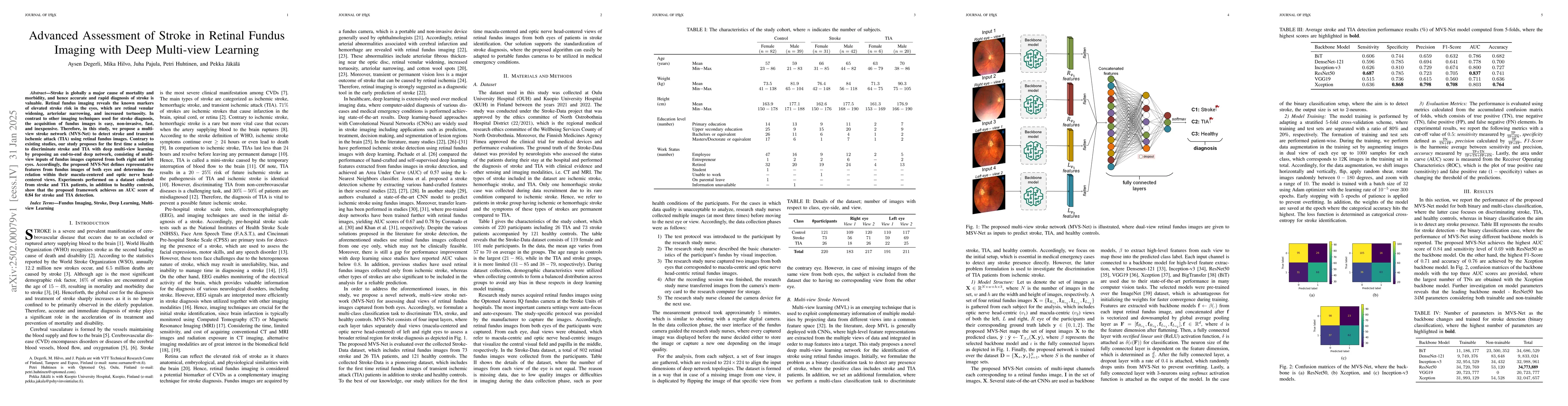
this study, we propose a multi-view stroke network (MVS-Net) to detect stroke and transient ischemic attack (TIA) using retinal fundus images. Contrary to existing studies, our study proposes for the first time a solution to discriminate stroke and TIA with deep multi-view learning by proposing an end-to-end deep network, consisting of multi-view inputs of fundus images captured from both right and left eyes. Accordingly, the proposed MVS-Net defines representative features from fundus images of both eyes and determines the relation within their macula-centered and optic nerve head-centered views. Experiments performed on a dataset collected from stroke and TIA patients, in addition to healthy controls, show that the proposed framework achieves an AUC score of 0.84 for stroke and TIA detection.
Key Findings, in focus
Seven facets of this paper, analysed and brought into focus by AI.
This research proposes a novel method for standardizing stroke diagnosis using retinal fundus images, which can be used even in remote areas with limited access to hospitals and neurologists.
- MVS-Net achieves an AUC score of 0.84 for stroke and TIA detection.
- ResNet50 backbone model achieves the highest sensitivity (0.69) and accuracy (0.76) in binary classification.
- Xception backbone model achieves the highest F1-score (0.71) and accuracy (0.76) in binary classification.
- Discriminating TIA from the rest of the dataset is identified as the most challenging task.
- Using multi-view retinal fundus images outperforms single-view approaches for stroke detection.
This research proposes a novel method for standardizing stroke diagnosis using retinal fundus images, which can be used even in remote areas with limited access to hospitals and neurologists. It highlights the potential of retinal fundus imaging with deep learning for low-cost stroke treatment improvement.
The paper introduces MVS-Net, a deep multi-view learning network that utilizes dual-view retinal fundus images for stroke detection, merging high-level features extracted from different views for prediction.
This study is the first to propose a multi-view deep learning network (MVS-Net) for discriminating stroke, TIA, and healthy controls using retinal fundus images from both eyes, outperforming single-view approaches.
- The Stroke-Data dataset is relatively small with 802 images from 220 participants, which may not be ideal for training deep learning models.
- No other publicly available multi-view retinal fundus image datasets exist for comparison.
- Explore larger and more diverse datasets to improve model performance and generalizability.
- Investigate the use of the proposed method for detecting other diseases that can be investigated with retinal fundus imaging and deep learning.
Authors
PDF Preview
Related Papers
No references found for this paper.
Discussion 0