Publication
Metrics
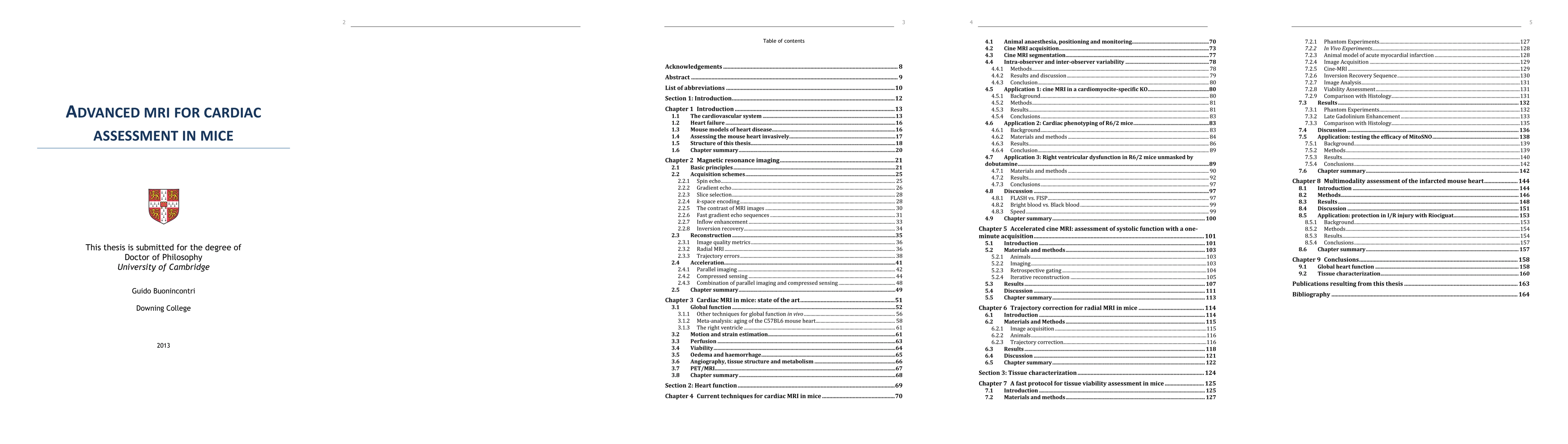
Paper Preview
Abstract
Heart failure is a leading cause of mortality in the Western world. The mouse is a widely used model for a number of diseases, induced by genetic modification or surgical intervention. When performing experiments in mice, in vivo magnetic resonance imaging (MRI) can be used to evaluate heart anatomy and function at multiple levels. Volumetric measurements of ventricle size at each phase of the heart cycle (obtained with cine MRI) can be used as a sensitive measure for heart failure. Cine MRI is applied here to genetic models of heart disease, including stress tests with pharmacological manipulation. As the long duration of the acquisition for functional assessment is a critical limitation, a new method to make cine MRI twelve times faster is presented and validated, maintaining standard accuracy. The method utilises a combination of compressed sensing and parallel imaging with a radial acquisition of k-space. Eddy-current induced artifacts, present when acquiring radially, are corrected retrospectively with a novel scheme. Tissue viability can be measured with late gadolinium enhancement (LGE) imaging, where a contrast agent is utilised. After injection, the agent rapidly washes out of healthy tissue, though has a slower kinetic in infarcted regions. A novel method to perform LGE efficiently is presented and validated against histology. This method is applied to the evaluation of a new treatment for infarction. Finally, a novel method to perform a multi-modal assessment of the mouse heart within a single exam is presented. The method includes cine and LGE MRI as well as a measure of mechanical strain in the myocardium with displacement encoding with stimulated echoes (DENSE) MRI and assessment of cellular metabolism with positron emission tomography (PET). This method is demonstrated in the evaluation of a new protective agent for infarction.
AI Key Findings
Get AI-generated insights about this paper's methodology, results, significance, and more — seven facets brought into focus.
Paper Details
Authors
PDF Preview
Key Terms
Related Papers
No references found for this paper.
Discussion 0