01
MethodologyHow they did it
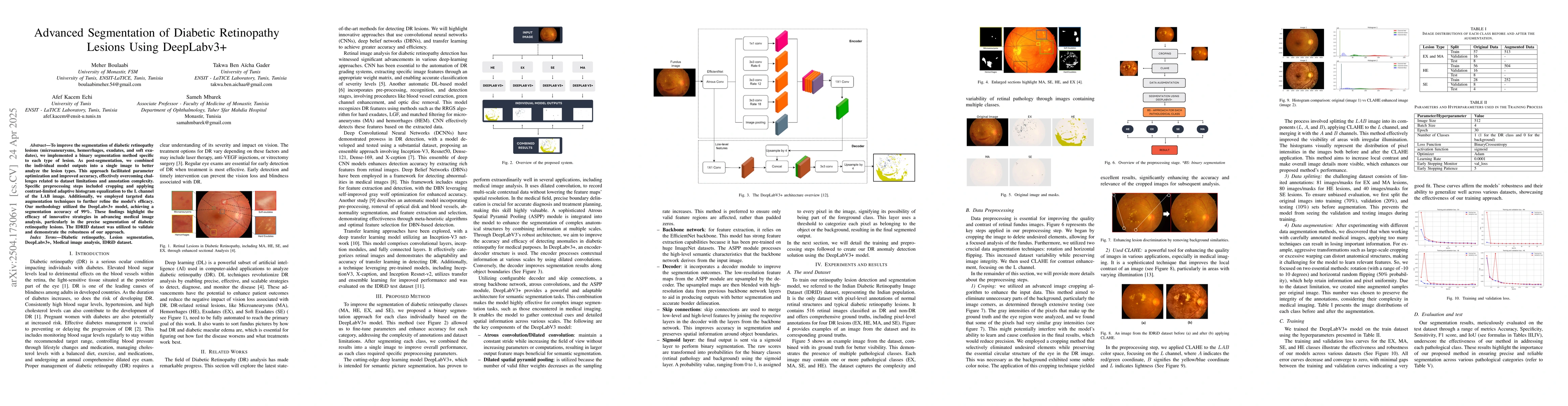
The research implemented a binary segmentation method specific to each type of diabetic retinopathy lesion (microaneurysms, hemorrhages, exudates, and soft exudates) using DeepLabv3+, combining individual model outputs into a single image for better analysis. Preprocessing steps included cropping and applying contrast-limited adaptive histogram equalization (CLAHE) to the L channel of the LAB image, along with targeted data augmentation techniques.
Discussion 0