Advances in Kidney Biopsy Lesion Assessment through Dense Instance Segmentation
Publication
Metrics
AI Quick Summary
This paper introduces DiffRegFormer, a dense instance segmentation model that efficiently classifies and segments over 500 anatomical objects within kidney biopsy regions-of-interest. The model demonstrates superior performance in detecting and segmenting glomeruli, tubuli, and arteries, achieving 52.1% average precision in detection and 46.8% in segmentation, and effectively reduces inter-observer variability in lesion assessment.
Paper Preview
Abstract
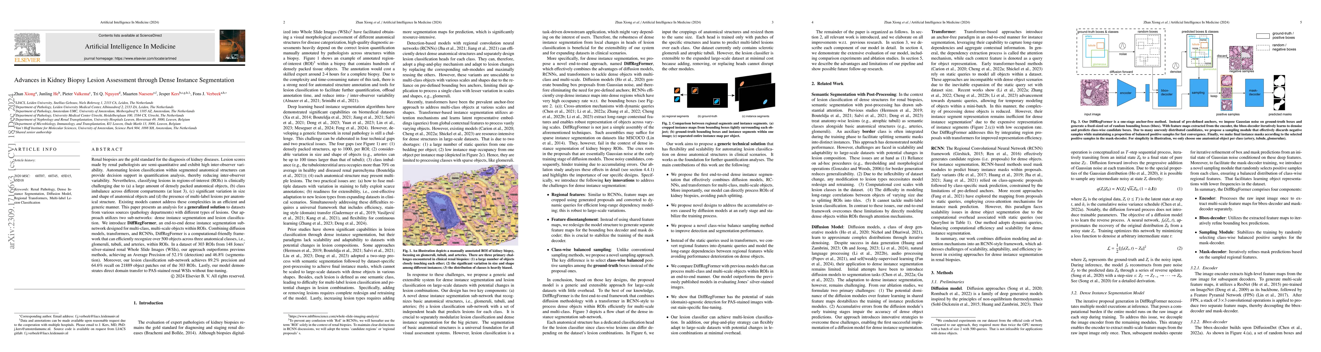
Renal biopsies are the gold standard for diagnosis of kidney diseases. Lesion scores made by renal pathologists are semi-quantitative and exhibit high inter-observer variability. Automating lesion classification within segmented anatomical structures can provide decision support in quantification analysis and reduce the inter-observer variability. Nevertheless, classifying lesions in regions-of-interest (ROIs) is clinically challenging due to (a) a large amount of densely packed anatomical objects (up to 1000), (b) class imbalance across different compartments (at least 3), (c) significant variation in object scales (i.e. sizes and shapes), and (d) the presence of multi-label lesions per anatomical structure. Existing models lack the capacity to address these complexities efficiently and generically. This paper presents \textbf{a generalized technical solution} for large-scale, multi-source datasets with diverse lesions. Our approach utilizes two sub-networks: dense instance segmentation and lesion classification. We introduce \textbf{DiffRegFormer}, an end-to-end dense instance segmentation model designed for multi-class, multi-scale objects within ROIs. Combining diffusion models, transformers, and RCNNs, DiffRegFormer efficiently recognizes over 500 objects across three anatomical classes (glomeruli, tubuli, arteries) within ROIs on a single NVIDIA GeForce RTX 3090 GPU. On a dataset of 303 ROIs (from 148 Jones' silver-stained renal WSIs), it outperforms state of art models, achieving AP of 52.1\% (detection) and 46.8\% (segmentation). Our lesion classification sub-network achieves 89.2\% precision and 64.6\% recall on 21889 object patches (from the 303 ROIs). Importantly, the model demonstrates direct domain transfer to PAS-stained WSIs without fine-tuning.
AI Key Findings
Get AI-generated insights about this paper's methodology, results, significance, and more — seven facets brought into focus.
Paper Details
Authors
PDF Preview
Key Terms
Related Papers
No references found for this paper.
Discussion 0