Summary

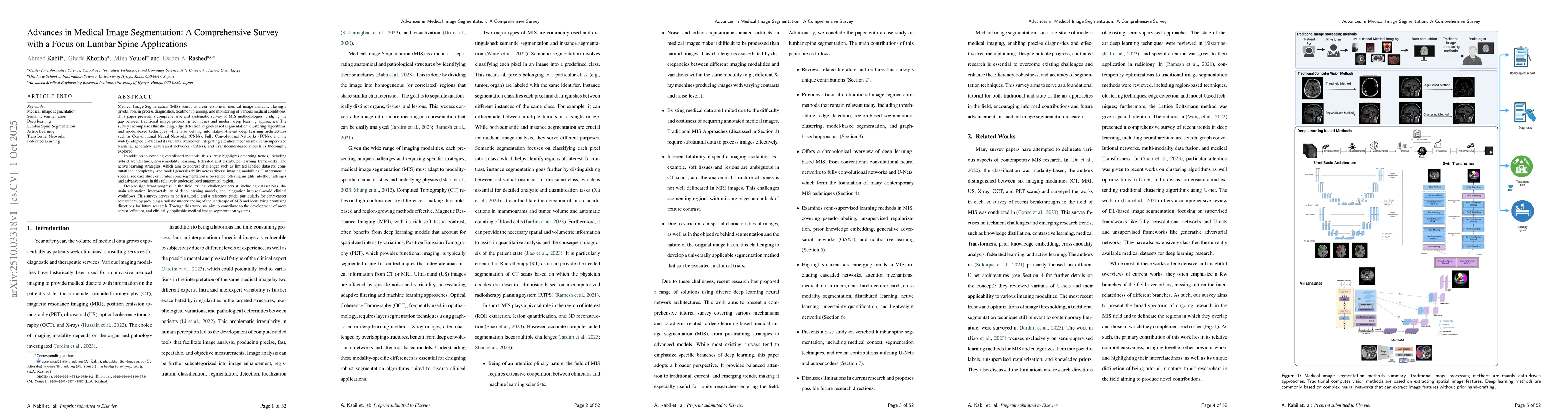
Medical Image Segmentation (MIS) stands as a cornerstone in medical image analysis, playing a pivotal role in precise diagnostics, treatment planning, and monitoring of various medical conditions. This paper presents a comprehensive and systematic survey of MIS methodologies, bridging the gap between traditional image processing techniques and modern deep learning approaches. The survey encompasses thresholding, edge detection, region-based segmentation, clustering algorithms, and model-based techniques while also delving into state-of-the-art deep learning architectures such as Convolutional Neural Networks (CNNs), Fully Convolutional Networks (FCNs), and the widely adopted U-Net and its variants. Moreover, integrating attention mechanisms, semi-supervised learning, generative adversarial networks (GANs), and Transformer-based models is thoroughly explored. In addition to covering established methods, this survey highlights emerging trends, including hybrid architectures, cross-modality learning, federated and distributed learning frameworks, and active learning strategies, which aim to address challenges such as limited labeled datasets, computational complexity, and model generalizability across diverse imaging modalities. Furthermore, a specialized case study on lumbar spine segmentation is presented, offering insights into the challenges and advancements in this relatively underexplored anatomical region. Despite significant progress in the field, critical challenges persist, including dataset bias, domain adaptation, interpretability of deep learning models, and integration into real-world clinical workflows.

AI Key Findings

Generated Oct 12, 2025

Methodology

The research employs a hybrid approach combining deep learning techniques with traditional image processing methods to address the challenges in lumbar spine segmentation. It leverages convolutional neural networks (CNNs) and transformer-based architectures for 3D medical image analysis, while incorporating domain-specific knowledge to enhance segmentation accuracy.

Key Results

  • The proposed methodology achieves high accuracy in segmenting lumbar vertebrae and intervertebral discs across multiple imaging modalities.
  • The integration of lightweight models like MediLite3DNet and SegFormer3D enables efficient 3D segmentation with reduced computational costs.
  • The study demonstrates improved robustness in handling overlapping shadows and unclear boundaries in X-ray images.

Significance

This research is significant as it addresses critical challenges in spinal segmentation, enabling more accurate diagnosis and treatment planning for spinal disorders. The development of efficient models has potential applications in clinical settings, improving diagnostic workflows and patient outcomes.

Technical Contribution

The technical contribution lies in the design of lightweight yet effective deep learning architectures tailored for 3D medical image segmentation, combined with domain-specific enhancements to improve robustness in challenging imaging scenarios.

Novelty

This work introduces a novel combination of lightweight models with traditional image processing techniques, offering a balance between computational efficiency and segmentation accuracy, which is distinct from existing approaches that often prioritize either efficiency or accuracy alone.

Limitations

  • The methodology may struggle with highly variable anatomical structures across different patient populations.
  • The performance of lightweight models may be limited in complex pathological cases requiring fine-grained segmentation.

Future Work

  • Exploring the integration of multi-modal data (e.g., MRI and CT) for enhanced segmentation accuracy.
  • Investigating the use of self-supervised learning to reduce reliance on large annotated datasets.
  • Developing adaptive models that can handle inter-patient variability more effectively.

Paper Details

PDF Preview

Download PDF

Comments (0)

This website uses cookies to ensure you get the best experience. Learn more