Authors
Summary
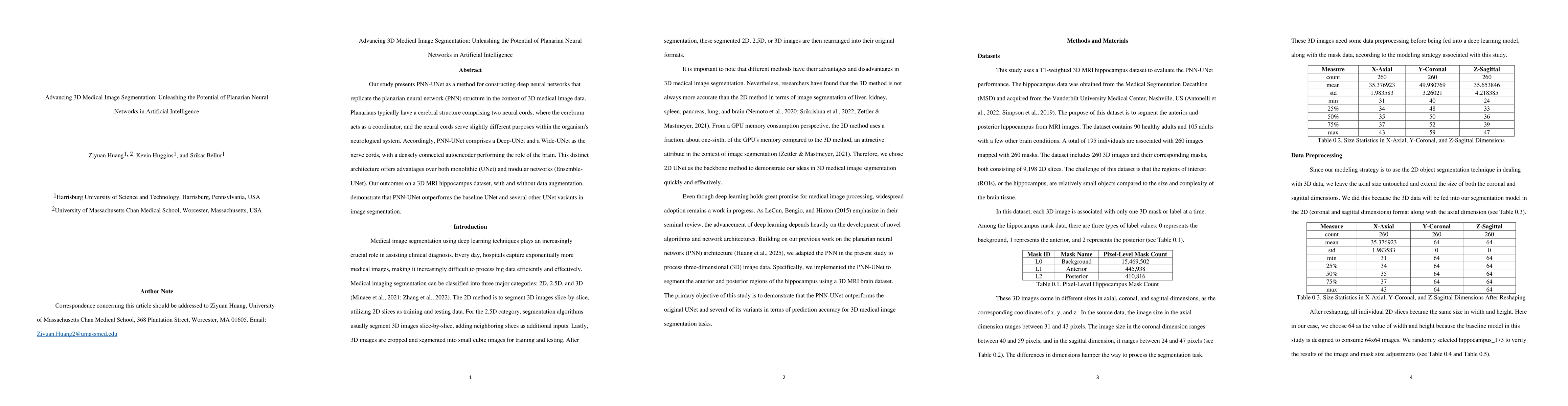
Our study presents PNN-UNet as a method for constructing deep neural networks that replicate the planarian neural network (PNN) structure in the context of 3D medical image data. Planarians typically have a cerebral structure comprising two neural cords, where the cerebrum acts as a coordinator, and the neural cords serve slightly different purposes within the organism's neurological system. Accordingly, PNN-UNet comprises a Deep-UNet and a Wide-UNet as the nerve cords, with a densely connected autoencoder performing the role of the brain. This distinct architecture offers advantages over both monolithic (UNet) and modular networks (Ensemble-UNet). Our outcomes on a 3D MRI hippocampus dataset, with and without data augmentation, demonstrate that PNN-UNet outperforms the baseline UNet and several other UNet variants in image segmentation.
AI Key Findings
Generated Sep 02, 2025
Methodology
The study introduces PNN-UNet, a deep neural network architecture inspired by the planarian neural network structure, comprising Deep-UNet and Wide-UNet as nerve cords and a densely connected autoencoder as the brain, for 3D medical image segmentation tasks.
Key Results
- PNN-UNet outperforms baseline models (Deep-UNet, Wide-UNet, Ensemble-Transfer, Ensemble-Retrain) in Dice and Jaccard scores for hippocampus segmentation in 3D MRI data, both with and without data augmentation.
- PNN-UNet demonstrates moderate improvements in sensitivity and specificity, though not statistically significant in all cases.
Significance
The research contributes to advancing 3D medical image segmentation by leveraging biological inspiration from planarian neural networks, potentially leading to more accurate and efficient AI models for medical imaging.
Technical Contribution
The PNN-UNet architecture, integrating deep and wide neural networks with a coordinating autoencoder, effectively improves segmentation performance by capitalizing on the strengths of both deep and wide architectures.
Novelty
This work is novel in its biologically inspired neural network design for medical image segmentation, combining deep and wide networks with an autoencoder to coordinate their functions, outperforming traditional UNet and ensemble methods.
Limitations
- The study did not observe significant improvements in sensitivity and specificity, which are crucial for assessing the model's ability to correctly identify hippocampus and background regions.
- The findings are based on a single dataset (3D MRI hippocampus), limiting the generalizability of results to other medical image types or anatomical structures.
Future Work
- Explore the application of PNN-UNet architecture to diverse medical image datasets for broader validation.
- Investigate the potential of PNN-UNet in semi-supervised, unsupervised, and reinforcement learning contexts to enhance its adaptability and efficiency.
Paper Details
PDF Preview
Citation Network
Current paper (gray), citations (green), references (blue)
Display is limited for performance on very large graphs.
Similar Papers
Found 4 papersMedical Image Segmentation with 3D Convolutional Neural Networks: A Survey
S Niyas, S J Pawan, M Anand Kumar et al.
3D TransUNet: Advancing Medical Image Segmentation through Vision Transformers
Xianhang Li, Lei Xing, Yuyin Zhou et al.
No citations found for this paper.
Comments (0)