Quick Answers
What methodology did the authors use?
The study integrates an attention-based 3D U-Net architecture with a tumor detection algorithm based on digital image processing techniques. The model is trained on the BraTS 2020 dataset using dice loss and evaluated with metrics like accuracy, Dice coefficient, IoU, sensitivity, and specificity. More in Methodology →
What are the key results?
The model achieved a Dice coefficient of 0.975, specificity of 0.988, and sensitivity of 0.995 — It outperformed related studies in segmentation accuracy More in Key Results →
Why is this work significant?
This research advances brain tumor segmentation in medical diagnostics, offering reliable and precise tools for clinicians to improve diagnosis and treatment planning. More in Significance →
What are the main limitations?
The model's performance may degrade with highly irregular tumor shapes — Class imbalance in training data remains a challenge despite preprocessing More in Limitations →
Abstract
In the realm of medical diagnostics, rapid advancements in Artificial Intelligence (AI) have significantly yielded remarkable improvements in brain tumor segmentation. Encoder-Decoder architectures, such as U-Net, have played a transformative role by effectively extracting meaningful representations in 3D brain tumor segmentation from Magnetic resonance imaging (MRI) scans. However, standard U-Net models encounter challenges in accurately delineating tumor regions, especially when dealing with irregular shapes and ambiguous boundaries. Additionally, training robust segmentation models on high-resolution MRI data, such as the BraTS datasets, necessitates high computational resources and often faces challenges associated with class imbalance. This study proposes the integration of the attention mechanism into the 3D U-Net model, enabling the model to capture intricate details and prioritize informative regions during the segmentation process. Additionally, a tumor detection algorithm based on digital image processing techniques is utilized to address the issue of imbalanced training data and mitigate bias. This study aims to enhance the performance of brain tumor segmentation, ultimately improving the reliability of diagnosis. The proposed model is thoroughly evaluated and assessed on the BraTS 2020 dataset using various performance metrics to accomplish this goal. The obtained results indicate that the model outperformed related studies, exhibiting dice of 0.975, specificity of 0.988, and sensitivity of 0.995, indicating the efficacy of the proposed model in improving brain tumor segmentation, offering valuable insights for reliable diagnosis in clinical settings.
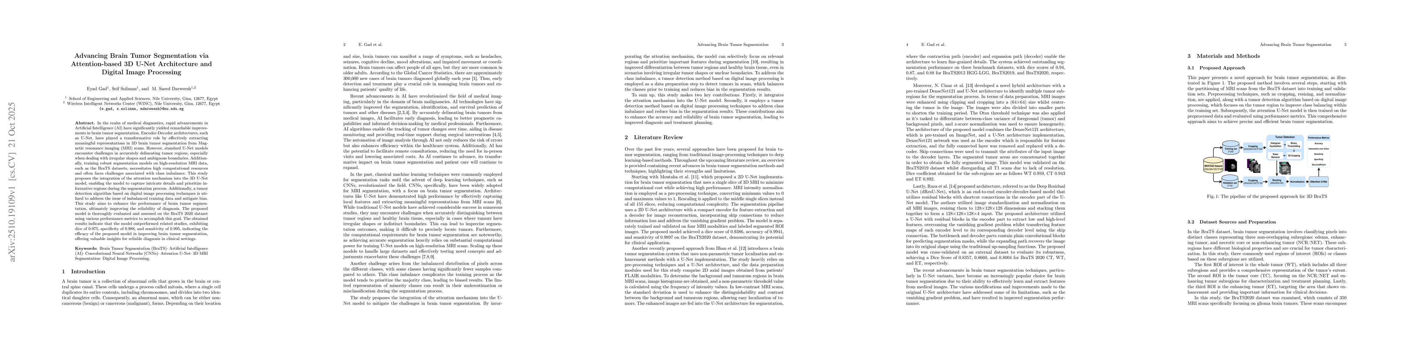
AI Key Findings
Generated Oct 24, 2025
Methodology — What approach did the authors take?
The study integrates an attention-based 3D U-Net architecture with a tumor detection algorithm based on digital image processing techniques. The model is trained on the BraTS 2020 dataset using dice loss and evaluated with metrics like accuracy, Dice coefficient, IoU, sensitivity, and specificity.
Key Results — What are the main findings?
- The model achieved a Dice coefficient of 0.975, specificity of 0.988, and sensitivity of 0.995
- It outperformed related studies in segmentation accuracy
- The attention mechanism improved focus on relevant tumor features
Significance — Why does this research matter?
This research advances brain tumor segmentation in medical diagnostics, offering reliable and precise tools for clinicians to improve diagnosis and treatment planning.
Technical Contribution — What is the technical contribution?
Development of an attention-based 3D U-Net with enhanced skip connections and a tumor detection algorithm for class balancing
Novelty — What is new about this work?
Integration of attention mechanisms into 3D U-Net and application of digital image processing for tumor detection, addressing class imbalance and improving segmentation accuracy
Limitations — What are the limitations of this study?
- The model's performance may degrade with highly irregular tumor shapes
- Class imbalance in training data remains a challenge despite preprocessing
Future Work — What did the authors propose for future work?
- Exploring multi-modal data fusion techniques for better tumor differentiation
- Investigating the model's performance on larger and more diverse datasets
Paper Details
How to Cite This Paper
@article{gad2025advancing,
title = {Advancing Brain Tumor Segmentation via Attention-based 3D U-Net
Architecture and Digital Image Processing},
author = {Gad, Eyad and Darweesh, M. Saeed and Soliman, Seif},
year = {2025},
eprint = {2510.19109},
archivePrefix = {arXiv},
primaryClass = {cs.CV},
doi = {10.1007/978-3-031-49333-1_18},
}
Gad, E., Darweesh, M., & Soliman, S. (2025). Advancing Brain Tumor Segmentation via Attention-based 3D U-Net
Architecture and Digital Image Processing. arXiv. https://doi.org/10.1007/978-3-031-49333-1_18
Gad, Eyad, et al. "Advancing Brain Tumor Segmentation via Attention-based 3D U-Net
Architecture and Digital Image Processing." arXiv, 2025, doi.org/10.1007/978-3-031-49333-1_18.
PDF Preview
Similar Papers
Found 4 papersMulticlass MRI Brain Tumor Segmentation using 3D Attention-based U-Net
Maryann M. Gitonga
3D U-Net Based Brain Tumor Segmentation and Survival Days Prediction
Feifan Wang, Liqin Zheng, Runzhou Jiang et al.
LATUP-Net: A Lightweight 3D Attention U-Net with Parallel Convolutions for Brain Tumor Segmentation
Ebtihal J. Alwadee, Xianfang Sun, Yipeng Qin et al.
U-Net-based architecture with attention mechanisms and Bayesian Optimization for brain tumor segmentation using MR images.
Ramalakshmi, K, Krishna Kumari, L
Comments (0)