Advancing EEG/MEG Source Imaging with Geometric-Informed Basis Functions
Publication
Metrics
AI Quick Summary
This paper introduces Brain Geometric-informed Basis Functions (GBFs) to improve EEG/MEG source imaging, demonstrating superior performance over traditional methods and existing ESI techniques through synthetic and real EEG data experiments, resulting in more robust and biologically interpretable brain activity reconstructions.
Paper Preview
Abstract
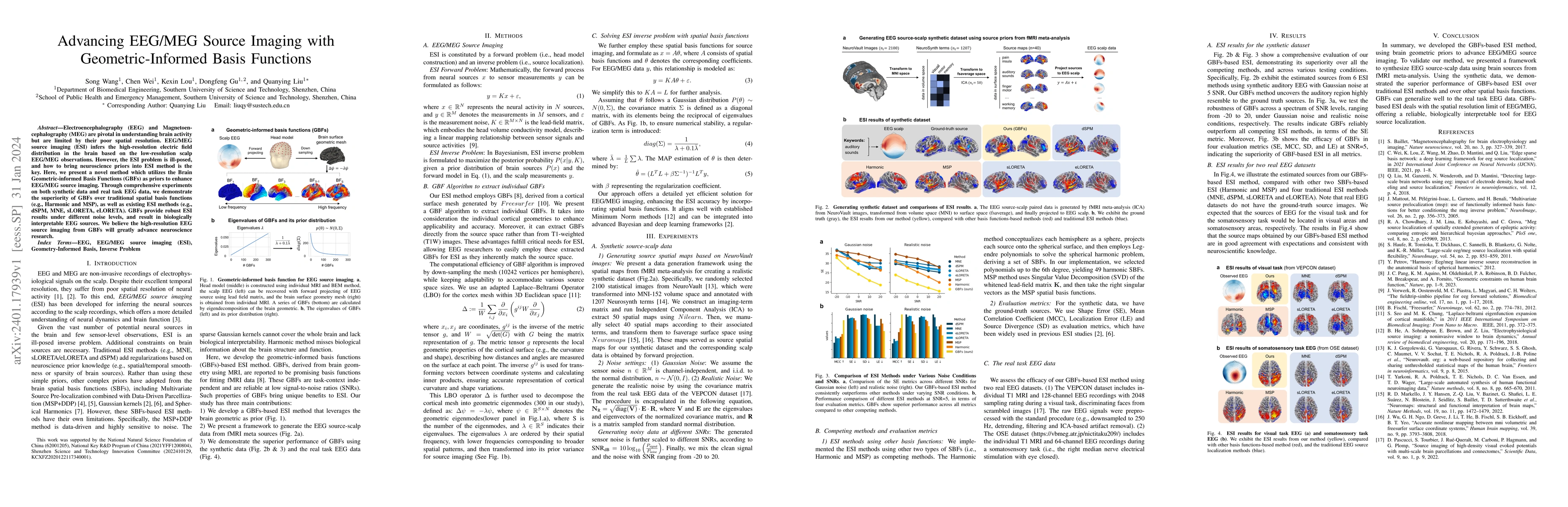
Electroencephalography (EEG) and Magnetoencephalography (MEG) are pivotal in understanding brain activity but are limited by their poor spatial resolution. EEG/MEG source imaging (ESI) infers the high-resolution electric field distribution in the brain based on the low-resolution scalp EEG/MEG observations. However, the ESI problem is ill-posed, and how to bring neuroscience priors into ESI method is the key. Here, we present a novel method which utilizes the Brain Geometric-informed Basis Functions (GBFs) as priors to enhance EEG/MEG source imaging. Through comprehensive experiments on both synthetic data and real task EEG data, we demonstrate the superiority of GBFs over traditional spatial basis functions (e.g., Harmonic and MSP), as well as existing ESI methods (e.g., dSPM, MNE, sLORETA, eLORETA). GBFs provide robust ESI results under different noise levels, and result in biologically interpretable EEG sources. We believe the high-resolution EEG source imaging from GBFs will greatly advance neuroscience research.
AI Key Findings
Get AI-generated insights about this paper's methodology, results, significance, and more — seven facets brought into focus.
Impact
Paper Details
Authors
PDF Preview
Key Terms
Citation Network
Current paper (gray), citations (green), references (blue)
Display is limited for performance on very large graphs.
Discussion 0