Summary

Grey matter loss in the hippocampus is a hallmark of neurobiological aging, yet understanding the corresponding changes in its functional connectivity remains limited. Seed-based functional connectivity (FC) analysis enables voxel-wise mapping of the hippocampus's synchronous activity with cortical regions, offering a window into functional reorganization during aging. In this study, we develop an interpretable deep learning framework to predict brain age from hippocampal FC using a three-dimensional convolutional neural network (3D CNN) combined with LayerCAM saliency mapping. This approach maps key hippocampal-cortical connections, particularly with the precuneus, cuneus, posterior cingulate cortex, parahippocampal cortex, left superior parietal lobule, and right superior temporal sulcus, that are highly sensitive to age. Critically, disaggregating anterior and posterior hippocampal FC reveals distinct mapping aligned with their known functional specializations. These findings provide new insights into the functional mechanisms of hippocampal aging and demonstrate the power of explainable deep learning to uncover biologically meaningful patterns in neuroimaging data.
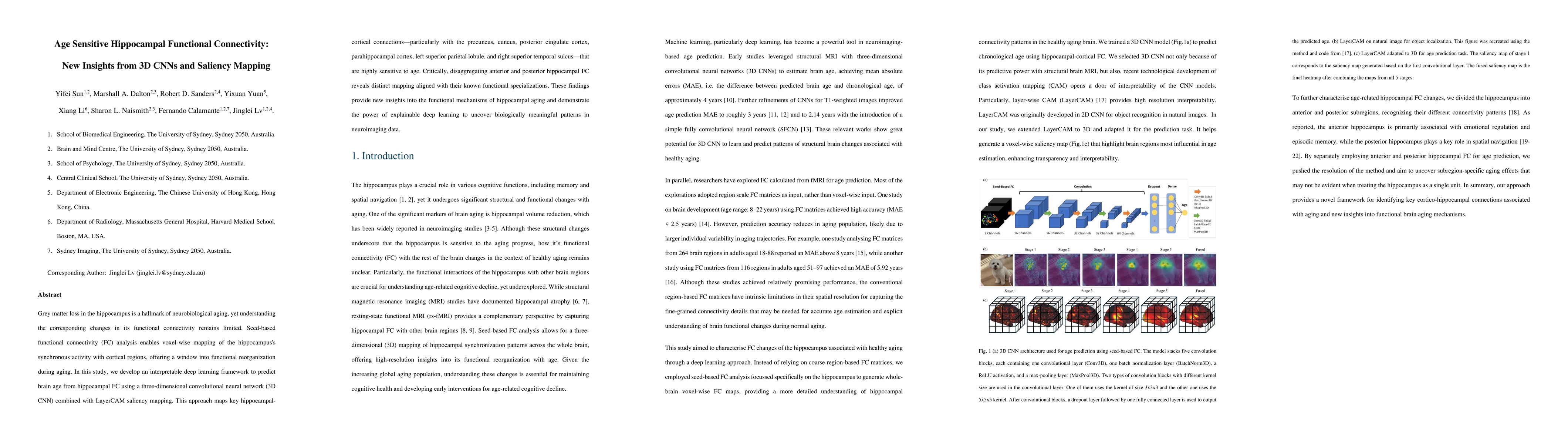
AI Key Findings

Generated Sep 03, 2025

Methodology

This study employs a 3D Convolutional Neural Network (CNN) model to predict chronological age based on voxel-wise cortico-hippocampal functional connectivity (FC), using LayerCAM for interpretable analysis. The model is pretrained on the Human Connectome Project Young Adult (HCP-Y) dataset and fine-tuned on the HCP-Aging (HCP-A) dataset for age prediction across a wide age range (22-90 years).

Key Results

  • The model achieved a Mean Absolute Error (MAE) of 2.99 years for young adults in the HCP-YA dataset, comparable to previous work using structural or diffusion MRI.
  • For the HCP-A dataset, the MAE increased to 7.30 years, outperforming a resting-state-only model from a recent study and narrowing the performance gap to just 1.65 years with finer-grained FC information.
  • Anterior hippocampal FC with the prefrontal cortex, anterior cingulate cortex, entorhinal cortex, and temporal pole showed stronger age-predictive salience, reflecting cognitive functions vulnerable to aging.
  • Posterior hippocampal FC with the precuneus, inferior parietal lobule, and superior temporal sulcus was predictive of age, potentially reflecting early functional changes associated with normal aging.
  • The study reveals distinct functional trajectories in normal aging for anterior and posterior hippocampal connectivity, which may link to the heterogeneity of age-related cognitive decline.

Significance

This research demonstrates the efficacy of deep learning in neuroimaging, offering new insights into the functional mechanisms of hippocampal aging and paving the way for more targeted investigations of brain regions related to normal aging.

Technical Contribution

The study introduces an interpretable deep learning method using 3D CNNs and LayerCAM saliency mapping to predict brain age from hippocampal FC, revealing key hippocampal-cortical connections sensitive to age.

Novelty

This work stands out by combining deep learning with LayerCAM for interpretable analysis of hippocampal FC changes in healthy aging, uncovering complex patterns not detectable by traditional linear regression methods.

Limitations

  • The study is limited to a healthy aging cohort and does not account for neurological conditions like dementia.
  • Expanding the model's focus across the entire lifespan, including infants and children, presents a challenging prospect due to substantial variability in brain development stages.

Future Work

  • Exploring the brain age gap and correlating it with cognitive measures, behavioral data, and genetic information could benefit precision medicine and personalized treatment.
  • Investigating alternative ROIs or incorporating additional relevant subcortical and cortical regions could potentially enhance predictive accuracy and provide a more comprehensive understanding of human brain functional changes during aging.

Paper Details

PDF Preview

Download PDF

Citation Network

Current paper (gray), citations (green), references (blue)

Display is limited for performance on very large graphs.

Current Paper
Citations
References
Click to view

No citations found for this paper.

Comments (0)

This website uses cookies to ensure you get the best experience. Learn more