The study employed a semi-supervised deep learning framework called Teacher-Teaching Assistant-Student (TTAS) to segment and quantify pleural effusion (PE) volume from contrast-enhanced CT scans. The model was trained on 100 manually annotated cases and validated on external datasets like PleThora12, comparing performance against state-of-the-art models such as nnU-Net and 3DU-Net.
AI-Assisted Pleural Effusion Volume Estimation from Contrast-Enhanced CT Images
Publication
Metrics
Quick Answers
What methodology did the authors use?
The study employed a semi-supervised deep learning framework called Teacher-Teaching Assistant-Student (TTAS) to segment and quantify pleural effusion (PE) volume from contrast-enhanced CT scans. The model was trained on 100 manually annotated cases and validated on external datasets like PleThora12, comparing performance against state-of-the-art models such as nnU-Net and 3DU-Net. More in Methodology →
What are the key results?
TTAS achieved a mean Dice score of 0.82, significantly outperforming nnU-Net's 0.73 (p < 0.0001). — TTAS demonstrated a four-fold lower mean Absolute Volume Difference (AbVD) of 6.49 mL compared to nnU-Net's 23.16 mL (p < 0.0001). More in Key Results →
Why is this work significant?
This research advances automated PE quantification in CT scans, enabling more accurate and efficient clinical management of pleural effusions. The TTAS framework's robust segmentation and volume estimation could improve diagnostic workflows and reduce manual effort in radiology. More in Significance →
What are the main limitations?
The study relied on a limited number of annotated training cases (100) and external validation datasets, which may affect generalizability. — Manual segmentation by radiologists showed moderate agreement (ICC 0.63), indicating potential variability in ground truth annotations. More in Limitations →
Abstract
Background: Pleural Effusions (PE) is a common finding in many different clinical conditions, but accurately measuring their volume from CT scans is challenging. Purpose: To improve PE segmentation and quantification for enhanced clinical management, we have developed and trained a semi-supervised deep learning framework on contrast-enhanced CT volumes. Materials and Methods: This retrospective study collected CT Pulmonary Angiogram (CTPA) data from internal and external datasets. A subset of 100 cases was manually annotated for model training, while the remaining cases were used for testing and validation. A novel semi-supervised deep learning framework, Teacher-Teaching Assistant-Student (TTAS), was developed and used to enable efficient training in non-segmented examinations. Segmentation performance was compared to that of state-of-the-art models. Results: 100 patients (mean age, 72 years, 28 [standard deviation]; 55 men) were included in the study. The TTAS model demonstrated superior segmentation performance compared to state-of-the-art models, achieving a mean Dice score of 0.82 (95% CI, 0.79 - 0.84) versus 0.73 for nnU-Net (p < 0.0001, Student's T test). Additionally, TTAS exhibited a four-fold lower mean Absolute Volume Difference (AbVD) of 6.49 mL (95% CI, 4.80 - 8.20) compared to nnU-Net's AbVD of 23.16 mL (p < 0.0001). Conclusion: The developed TTAS framework offered superior PE segmentation, aiding accurate volume determination from CT scans.
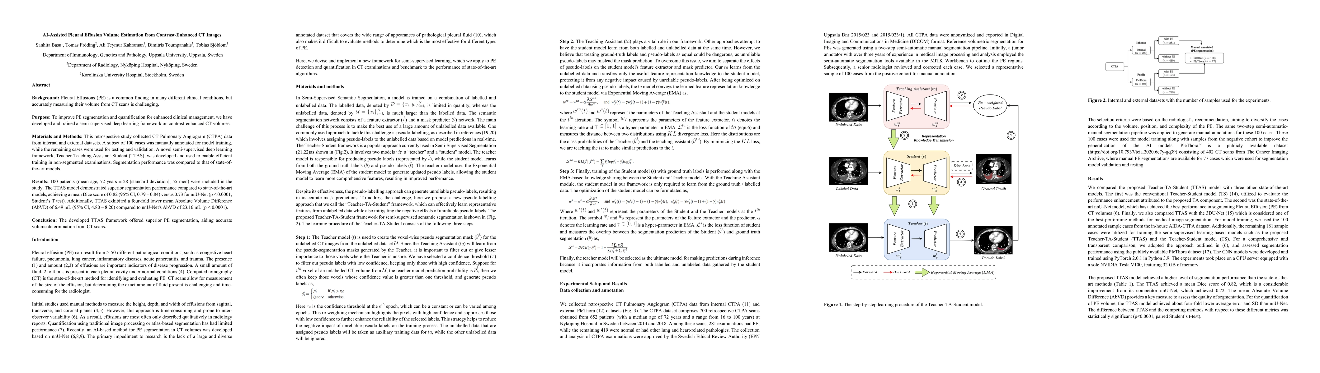
Key Findings, in focus
Seven facets of this paper, analysed and brought into focus by AI.
This research advances automated PE quantification in CT scans, enabling more accurate and efficient clinical management of pleural effusions.
- TTAS achieved a mean Dice score of 0.82, significantly outperforming nnU-Net's 0.73 (p < 0.0001).
- TTAS demonstrated a four-fold lower mean Absolute Volume Difference (AbVD) of 6.49 mL compared to nnU-Net's 23.16 mL (p < 0.0001).
- TTAS showed superior segmentation accuracy for large PE volumes (>200 mL) with a Dice score of 0.82 and reduced false negatives/false positives compared to existing methods.
This research advances automated PE quantification in CT scans, enabling more accurate and efficient clinical management of pleural effusions. The TTAS framework's robust segmentation and volume estimation could improve diagnostic workflows and reduce manual effort in radiology.
The TTAS framework introduces a novel semi-supervised learning approach that combines accurate labels with pseudo-labels for feature learning, while using only accurate labels for mask prediction, improving segmentation accuracy and volume estimation.
The TTAS model distinguishes itself by incorporating a Teaching Assistant component that enhances semi-supervised learning, reducing false positives and improving segmentation precision compared to conventional Teacher-Student models.
- The study relied on a limited number of annotated training cases (100) and external validation datasets, which may affect generalizability.
- Manual segmentation by radiologists showed moderate agreement (ICC 0.63), indicating potential variability in ground truth annotations.
- Expanding the dataset to include more diverse patient populations and CT scanners from different manufacturers.
- Developing a web-based interface for integrating the volume quantification model with PACS systems for clinical use.
Discussion 0