This paper explores the use of AI-based machine learning for analyzing super-resolution microscopy data to discover new biological insights without relying on known ground truth, employing weakly supervised learning techniques to reveal nanoscale architecture of cellular structures.
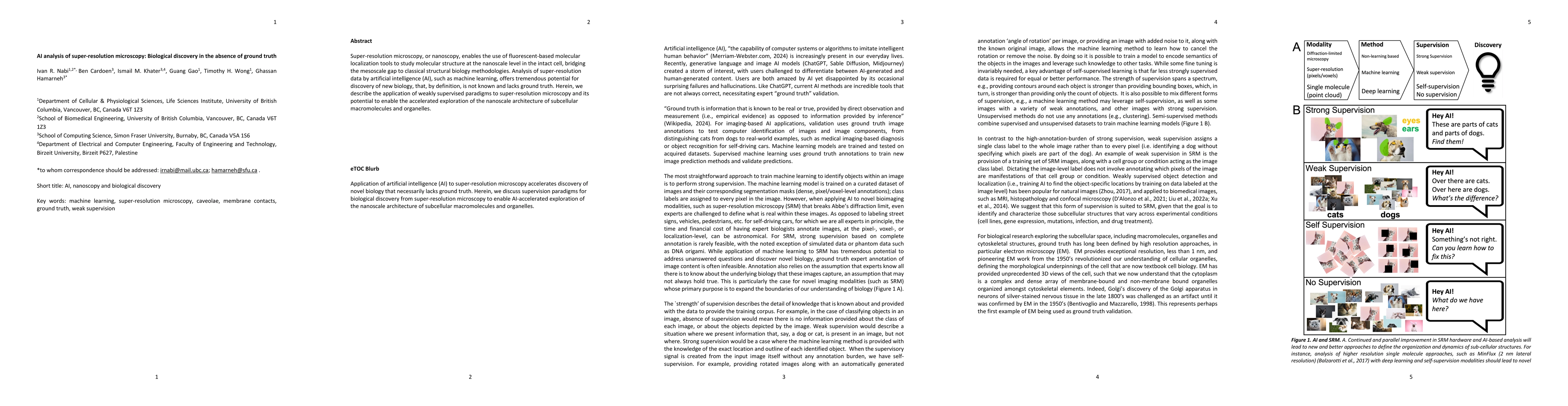
Super-resolution microscopy, or nanoscopy, enables the use of fluorescent-based molecular localization tools to study molecular structure at the nanoscale level in the intact cell, bridging the mesoscale gap to classical structural biology methodologies. Analysis of super-resolution data by artificial intelligence (AI), such as machine learning, offers tremendous potential for discovery of new biology, that, by definition, is not known and lacks ground truth. Herein, we describe the application of weakly supervised paradigms to super-resolution microscopy and its potential to enable the accelerated exploration of the nanoscale architecture of subcellular macromolecules and organelles.
Get AI-generated insights about this paper's methodology, results, and significance.
Current paper (gray), citations (green), references (blue)
Mario Krenn, Sören Arlt, Carla Rodríguez et al.
Serdar Kadioglu, Filip Michalsky
Title | Authors | Year | Actions |
---|---|---|---|
Scaffolds and the scaffolding domain: an alternative paradigm for caveolin-1 signaling
|
John E Lim, Pascal Bernatchez, Ivan R. Nabi
|
2024
|
View Paper |
Title | Authors | Year | Actions |
---|---|---|---|
Computational drug development for membrane protein targets
|
Haijian Li, Xiaolin Sun, Wenqiang Cui, Marc Xu, Junlin Dong, Babatunde E. Ekundayo, D. Ni, Zhili Rao, Liwei Guo, Henning Stahlberg, Shuguang Yuan, Horst Vogel
|
2024
|
View Paper |
XAI for All: Can Large Language Models Simplify Explainable AI?
|
Philip Mavrepis, Georgios Makridis, G. Fatouros, Vasileios Koukos, Maria Margarita Separdani, D. Kyriazis
|
2024
|
View Paper |
The rise of data-driven microscopy powered by machine learning.
|
Leonor Morgado, Estibaliz Gómez-de-Mariscal, H. Heil, Ricardo Henriques
|
2024
|
View Paper |
Harnessing eXplainable artificial intelligence for feature selection in time series energy forecasting: A comparative analysis of Grad-CAM and SHAP
|
Corne van Zyl, Xianming Ye, R. Naidoo
|
2024
|
View Paper |
Membrane contact site detection (MCS-DETECT) reveals dual control of rough mitochondria–ER contacts
|
Ben Cardoen, Kurt R Vandevoorde, G. Gao, Milene Ortiz-Silva, P. Alan, William Liu, Ellie Tiliakou, A. Vogl, Ghassan Hamarneh, I. Nabi
|
2023
|
View Paper |
Advanced optical imaging for the rational design of nanomedicines.
|
Ana Ortiz-Perez, Miao Zhang, Laurence W Fitzpatrick, Cristina Izquierdo-Lozano, L. Albertazzi
|
2023
|
View Paper |
Live-cell imaging in the deep learning era.
|
Joanna W. Pylvänäinen, Estibaliz Gómez-de-Mariscal, Ricardo Henriques, Guillaume Jacquemet
|
2023
|
View Paper |
How to estimate carbon footprint when training deep learning models? A guide and review
|
Lucía Bouza, Aurélie Bugeau, Loïc Lannelongue
|
2023
|
View Paper |
DataCurator.jl: efficient, portable and reproducible validation, curation and transformation of large heterogeneous datasets using human-readable recipes compiled into machine-verifiable templates
|
Ben Cardoen, Hanene Ben Yedder, Sieun Lee, I. Nabi, Ghassan Hamarneh
|
2023
|
View Paper |
SEMORE: SEgmentation and MORphological fingErprinting by machine learning automates super-resolution data analysis
|
Steen Bender, M. W. Dreisler, Min Zhang, Jacob Kæstel-Hansen, N. Hatzakis
|
2023
|
View Paper |
Machine-learning-powered extraction of molecular diffusivity from single-molecule images for super-resolution mapping
|
Ha H. Park, Bowen Wang, Suhong Moon, Tyler Jepson, Ke Xu
|
2023
|
View Paper |
Roadmap on Deep Learning for Microscopy.
|
Giovanni Volpe, Carolina Wahlby, L. Tian, M. Hecht, A. Yakimovich, Kristina Monakhova, L. Waller, I. Sbalzarini, Christopher A. Metzler, Mingyang Xie, Kevin Zhang, Isaac C. D. Lenton, H. Rubinsztein-Dunlop, D. Brunner, Bijie Bai, Aydogan Ozcan, Daniel Midtvedt, Hao Wang, Natavsa Sladoje, Joakim Lindblad, Jason T. Smith, M. Ochoa, Margarida M Barroso, X. Intes, Tong Qiu, Li-Yu Yu, Sixian You, Yongtao Liu, M. Ziatdinov, S. Kalinin, Arlo Sheridan, U. Manor, E. Nehme, Ofri Goldenberg, Y. Shechtman, H. Moberg, C. Langhammer, Barbora vSpavckov'a, Saga Helgadottir, Benjamin Midtvedt, Aykut Argun, T. Thalheim, F. Cichos, Stefano Bo, Lars Hubatsch, Jesús Pineda, Carlo Manzo, Harshith Bachimanchi, Erik Selander, A. Homs-Corbera, M. Franzl, Kevin de Haan, Y. Rivenson, Zofia Korczak, C. Adiels, M. Mijalkov, D'aniel Ver'eb, Yu-Wei Chang, J. Pereira, Damian J. Matuszewski, G. Kylberg, I. Sintorn, Juan C. Caicedo, B. Cimini, M. Bell, Bruno M. Saraiva, Guillaume Jacquemet, Ricardo Henriques, W. Ouyang, Trang Le, Estibaliz G'omez-de-Mariscal, D. Sage, A. Muñoz-Barrutia, Ebba Josefson Lindqvist, Johanna Bergman
|
2023
|
View Paper |
Community-developed checklists for publishing images and image analyses
|
C. Schmied, Michael S Nelson, S. Avilov, G. Bakker, Cristina Bertocchi, Johanna Bischof, U. Boehm, J. Brocher, Mariana T Carvalho, C. Chiritescu, J. Christopher, B. Cimini, Eduardo Conde-Sousa, M. Ebner, R. Ecker, K. Eliceiri, J. Fernandez-Rodriguez, Nathalie Gaudreault, L. Gelman, D. Grunwald, Tingting Gu, Nadia Halidi, Mathias Hammer, Matthew J. Hartley, M. Held, Florian Jug, V. Kapoor, Aysel Koksoy, J. Lacoste, Sylvia Le D'ev'edec, Sylvie Le Guyader, Peng Liu, Gabriel G Martins, Aastha Mathur, K. Miura, P. Llopis, R. Nitschke, A. North, A. Parslow, Alex L. Payne-Dwyer, Laure Plantard, Ali Rizwan, B. Schroth‐Diez, Lucas Schutz, Ryan T. Scott, A. Seitz, O. Selchow, V. Sharma, M. Spitaler, Sathya Srinivasan, Caterina Strambio de Castillia, D. Taatjes, C. Tischer, Helena Klara Jambor Fondazione Human Technopole, Milano, Italy, Centre for Bioimage Analysis, Embl, Heidelberg, Germany NCT-UCC, M. Dresden, Dresden., H Germany
|
2023
|
View Paper |
Clathrin coats partially preassemble and subsequently bend during endocytosis
|
M. Mund, Aline Tschanz, Yu-Le Wu, F. Frey, J. Mehl, M. Kaksonen, Ori Avinoam, U. Schwarz, J. Ries
|
2023
|
View Paper |
Recent development of computational cluster analysis methods for single-molecule localization microscopy images
|
Y. Hyun, Doory Kim
|
2023
|
View Paper |
Survey of Explainable AI Techniques in Healthcare
|
A. Chaddad, Jihao Peng, Jian Xu, A. Bouridane
|
2023
|
View Paper |
Caveolin-1 dolines form a distinct and rapid caveolae-independent mechanoadaptation system
|
Fidel-Nicolás Lolo, Nikhil Walani, Eric Seemann, D. Zalvidea, D. M. Pavón, G. Cojoc, M. Zamai, Christine Viaris de Lesegno, Fernando Martínez de Benito, M. Sánchez-Álvarez, Juan J. Uriarte, A. Echarri, Daniel Jimenez-Carretero, Joan-Carles Escolano, S. Sánchez, V. Caiolfa, D. Navajas, X. Trepat, J. Guck, C. Lamaze, Pere Roca-Cusachs, M. Kessels, B. Qualmann, M. Arroyo, M. A. del Pozo
|
2022
|
View Paper |
Evaluating the Clinical Utility of Artificial Intelligence Assistance and its Explanation on Glioma Grading Task
|
W. Jin, M. Fatehi, R. Guo, G. Hamarneh
|
2022
|
View Paper |
Rationalized deep learning super-resolution microscopy for sustained live imaging of rapid subcellular processes
|
Chang Qiao, Di Li, Yong Liu, Siwei Zhang, Kan Liu, Chong Liu, Yuting Guo, Tao Jiang, Chuyu Fang, Nan Li, Yunmin Zeng, Kangmin He, Xueliang Zhu, J. Lippincott-Schwartz, Qionghai Dai, Dong Li
|
2022
|
View Paper |
Explainable Convolutional Neural Networks: A Taxonomy, Review, and Future Directions
|
Rami Ibrahim, M. O. Shafiq
|
2022
|
View Paper |
Direct observation of motor protein stepping in living cells using MINFLUX
|
T. Deguchi, Malina K. Iwanski, Eva-Maria Schentarra, Christopher Heidebrecht, Lisa Schmidt, J. Heck, T. Weihs, Sebastian Schnorrenberg, P. Hoess, Sheng Liu, Veronika Chevyreva, K. Noh, L. Kapitein, J. Ries
|
2022
|
View Paper |
Precise measurement of nanoscopic septin ring structures with deep learning-assisted quantitative superresolution microscopy
|
Amin Zehtabian, Paul Markus Müller, Maximilian Goisser, Leon Obendorf, Lea Jänisch, Nadja Hümpfer, J. Rentsch, Helge Ewers
|
2022
|
View Paper |
Self-supervised deep learning encodes high-resolution features of protein subcellular localization
|
Hirofumi Kobayashi, Keith C. Cheveralls, M. Leonetti, Loic A. Royer
|
2022
|
View Paper |
Guidelines and evaluation of clinical explainable AI in medical image analysis
|
Weina Jin, Xiaoxiao Li, M. Fatehi, G. Hamarneh
|
2022
|
View Paper |
DynaMorph: self-supervised learning of morphodynamic states of live cells
|
Zhenqin Wu, Bryant B. Chhun, Galina Popova, Syuan-Ming Guo, Chang N. Kim, Li-Hao Yeh, Tomasz J. Nowakowski, James Zou, Shalin B. Mehta
|
2022
|
View Paper |
Super-Resolution Microscopy for Structural Cell Biology.
|
Sheng Liu, P. Hoess, J. Ries
|
2022
|
View Paper |
Precise measurement of nanoscopic septin ring structures in deep learning-assisted quantitative superresolution microscopy
|
Amin Zehtabian, P. M. Müller, Maximilian Goisser, Leon Obendorf, Lea Jänisch, Nadja Hümpfer, J. Rentsch, H. Ewers
|
2021
|
View Paper |
Is the replication crisis a base-rate fallacy?
|
B. Autzen
|
2021
|
View Paper |
Content-aware frame interpolation (CAFI): deep learning-based temporal super-resolution for fast bioimaging
|
Martin Priessner, David C. A. Gaboriau, Arlo Sheridan, Tchern Lenn, Jonathan R. Chubb, U. Manor, R. Vilar, Romain F. Laine
|
2021
|
View Paper |
Maximum-likelihood model fitting for quantitative analysis of SMLM data
|
Yu-Le Wu, P. Hoess, Aline Tschanz, U. Matti, M. Mund, J. Ries
|
2021
|
View Paper |
Highly accurate protein structure prediction with AlphaFold
|
J. Jumper, Richard Evans, A. Pritzel, Tim Green, Michael Figurnov, O. Ronneberger, Kathryn Tunyasuvunakool, Russ Bates, Augustin Žídek, Anna Potapenko, Alex Bridgland, Clemens Meyer, Simon A A Kohl, Andy Ballard, A. Cowie, Bernardino Romera-Paredes, Stanislav Nikolov, Rishub Jain, J. Adler, T. Back, Stig Petersen, D. Reiman, Ellen Clancy, Michal Zielinski, Martin Steinegger, Michalina Pacholska, Tamas Berghammer, S. Bodenstein, David Silver, O. Vinyals, A. Senior, K. Kavukcuoglu, Pushmeet Kohli, D. Hassabis
|
2021
|
View Paper |
Advancing biological super-resolution microscopy through deep learning: a brief review
|
Tianjie Yang, Yaoru Luo, Wei Ji, Ge Yang
|
2021
|
View Paper |
Can contrastive learning avoid shortcut solutions?
|
Joshua Robinson, Li Sun, Ke Yu, K. Batmanghelich, S. Jegelka, S. Sra
|
2021
|
View Paper |
Deep learning connects DNA traces to transcription to reveal predictive features beyond enhancer–promoter contact
|
Aparna R. Rajpurkar, Leslie J. Mateo, Sedona E. Murphy, A. Boettiger
|
2021
|
View Paper |
Weakly Supervised Deep Learning Approach to Breast MRI Assessment.
|
Michael Z. Liu, Cara Swintelski, Shawn H. Sun, Maham Siddique, E. Desperito, S. Jambawalikar, Richard S. Ha
|
2021
|
View Paper |
A survey on applications of deep learning in microscopy image analysis
|
Zhichao Liu, Luhong Jin, Jincheng Chen, Qiuyu Fang, S. Ablameyko, Zhaozheng Yin, Yingke Xu
|
2021
|
View Paper |
REMBI: Recommended Metadata for Biological Images—enabling reuse of microscopy data in biology
|
Ugis Sarkans, Wah Chiu, L. Collinson, M. Darrow, J. Ellenberg, D. Grunwald, J. Hériché, Andrii Iudin, G. Martins, T. Meehan, Kedar Narayan, A. Patwardhan, M. Russell, H. Saibil, C. Strambio‐De‐Castillia, J. Swedlow, C. Tischer, V. Uhlmann, P. Verkade, Mary Barlow, O. Bayraktar, E. Birney, Cesare Catavitello, C. Cawthorne, Stephan Wagner-Conrad, Elizabeth Duke, P. Paul-Gilloteaux, Emmanuel Gustin, M. Harkiolaki, P. Kankaanpää, T. Lemberger, J. Mcentyre, Josh Moore, A. Nicholls, Shuichi Onami, H. Parkinson, M. Parsons, M. Romanchikova, Nicholas J Sofroniew, J. Swoger, Nadine Utz, L. M. Voortman, Frances Wong, Peijun Zhang, G. Kleywegt, A. Brazma
|
2021
|
View Paper |
DEEMD: Drug Efficacy Estimation Against SARS-CoV-2 Based on Cell Morphology With Deep Multiple Instance Learning
|
M. Saberian, Kathleen P. Moriarty, A. Olmstead, Christian Hallgrimson, Franccois Jean, I. Nabi, Maxwell W. Libbrecht, Ghassan Hamarneh
|
2021
|
View Paper |
Mitochondria-rough-ER contacts in the liver regulate systemic lipid homeostasis.
|
I. Anastasia, Nicolò Ilacqua, A. Raimondi, Philippe Lemieux, Rana Ghandehari-Alavijeh, Guilhem Faure, S. Mekhedov, K. Williams, F. Caicci, Giorgio Valle, M. Giacomello, Ariel D. Quiroga, R. Lehner, M. Miksis, K. Tóth, Thomas Q. de Aguiar Vallim, E. Koonin, L. Scorrano, L. Pellegrini
|
2021
|
View Paper |
MINFLUX nanometer-scale 3D imaging and microsecond-range tracking on a common fluorescence microscope
|
R. Schmidt, T. Weihs, C. Wurm, Isabelle Jansen, Jasmin Rehman, S. Sahl, S. Hell
|
2021
|
View Paper |
Semantic segmentation of reflectance confocal microscopy mosaics of pigmented lesions using weak labels
|
Marissa D'Alonzo, A. Bozkurt, C. Alessi-Fox, M. Gill, D. Brooks, M. Rajadhyaksha, K. Kose, Jennifer G. Dy
|
2021
|
View Paper |
Off-the-shelf deep learning is not enough, and requires parsimony, Bayesianity, and causality
|
R. Vasudevan, M. Ziatdinov, L. Vlček, Sergei V. Kalinin
|
2021
|
View Paper |
A global view of standards for open image data formats and repositories
|
J. Swedlow, P. Kankaanpää, Ugis Sarkans, W. Goscinski, Graham Galloway, Leonel Malacrida, Ryan P. Sullivan, Steffen Härtel, C. Brown, C. Wood, Antje Keppler, Federica Paina, B. Loos, S. Zullino, D. Longo, S. Aime, Shuichi Onami
|
2020
|
View Paper |
SPECHT: Self-tuning Plausibility based object detection Enables quantification of Conflict in Heterogeneous multi-scale microscopy
|
Ben Cardoen, Timothy H. Wong, P. Alan, Sieun Lee, J. Matsubara, I. Nabi, G. Hamarneh
|
2020
|
View Paper |
Structures and distributions of SARS-CoV-2 spike proteins on intact virions
|
Zunlong Ke, J. Otón, K. Qu, M. Cortese, V. Zila, Lesley McKeane, T. Nakane, J. Zivanov, C. Neufeldt, Berati Cerikan, John M. Lu, J. Peukes, X. Xiong, H. Kräusslich, S. Scheres, R. Bartenschlager, J. Briggs
|
2020
|
View Paper |
Structure and assembly of CAV1 8S complexes revealed by single particle electron microscopy
|
Bing Han, J. Porta, Jessica L. Hanks, Y. Peskova, E. Binshtein, K. Dryden, D. P. Claxton, H. Mchaourab, E. Karakas, M. Ohi, A. Kenworthy
|
2020
|
View Paper |
Art and Science of the Cellular Mesoscale.
|
D. Goodsell, A. Olson, Stefano Forli
|
2020
|
View Paper |
Super resolution microscopy and deep learning identify Zika virus reorganization of the endoplasmic reticulum
|
Rory K. M. Long, Kathleen P. Moriarty, Ben Cardoen, G. Gao, A. Vogl, F. Jean, G. Hamarneh, I. Nabi
|
2020
|
View Paper |
Robust classification of cell cycle phase and biological feature extraction by image-based deep learning
|
Y. Nagao, Mika Sakamoto, Takumi Chinen, Y. Okada, Daisuke Takao
|
2020
|
View Paper |
ERGO: Efficient Recurrent Graph Optimized Emitter Density Estimation in Single Molecule Localization Microscopy
|
Ben Cardoen, Hanene Ben Yedder, Anmol Sharma, K. Chou, I. Nabi, G. Hamarneh
|
2019
|
View Paper |
Importance of potassium ions for ribosome structure and function revealed by long-wavelength X-ray diffraction
|
A. Rozov, I. Khusainov, K. el Omari, R. Duman, V. Mykhaylyk, M. Yusupov, E. Westhof, A. Wagner, G. Yusupova
|
2019
|
View Paper |
Pathologist-level interpretable whole-slide cancer diagnosis with deep learning
|
Zizhao Zhang, Pingjun Chen, M. McGough, Fuyong Xing, Chunbao Wang, M. Bui, Yuanpu Xie, Manish Sapkota, Lei Cui, J. Dhillon, Nazeel Ahmad, F. Khalil, S. Dickinson, Xiaoshuang Shi, Fujun Liu, H. Su, Jinzheng Cai, Lin Yang
|
2019
|
View Paper |
Coming together to define membrane contact sites
|
L. Scorrano, M. D. De Matteis, S. Emr, F. Giordano, G. Hajnóczky, Benoît Kornmann, L. Lackner, T. Levine, L. Pellegrini, K. Reinisch, R. Rizzuto, T. Simmen, H. Stenmark, C. Ungermann, M. Schuldiner
|
2019
|
View Paper |
Caveolae and scaffold detection from single molecule localization microscopy data using deep learning
|
Ismail M. Khater, Stephane T Aroca-Ouellette, Fanrui Meng, I. Nabi, G. Hamarneh
|
2019
|
View Paper |
Machine-learning for cluster analysis of localization microscopy data
|
David J. Williamson, G. Burn, J. Griffié, D. Davis, D. Owen
|
2018
|
View Paper |
Super-resolution modularity analysis shows polyhedral caveolin-1 oligomers combine to form scaffolds and caveolae
|
Ismail M. Khater, Qian T. Liu, K. Chou, G. Hamarneh, I. Nabi
|
2018
|
View Paper |
Deep‐learning‐based, computer‐aided classifier developed with a small dataset of clinical images surpasses board‐certified dermatologists in skin tumour diagnosis
|
Y. Fujisawa, Y. Otomo, Y. Ogata, Yasuhiro Nakamura, R. Fujita, Y. Ishitsuka, R. Watanabe, N. Okiyama, K. Ohara, Manabu Fujimoto
|
2018
|
View Paper |
Generative Adversarial Network in Medical Imaging: A Review
|
Xin Yi, Ekta Walia, P. Babyn
|
2018
|
View Paper |
Super Resolution Network Analysis Defines the Molecular Architecture of Caveolae and Caveolin-1 Scaffolds
|
Ismail M. Khater, Fanrui Meng, Timothy H. Wong, I. Nabi, G. Hamarneh
|
2018
|
View Paper |
Continual Lifelong Learning with Neural Networks: A Review
|
G. I. Parisi, Ronald Kemker, Jose L. Part, Christopher Kanan, Stefan Wermter
|
2018
|
View Paper |
Structured illumination microscopy combined with machine learning enables the high throughput analysis and classification of virus structure
|
Romain F. Laine, Gemma Goodfellow, Laurence J Young, J. Travers, Danielle Carroll, O. Dibben, H. Bright, C. Kaminski
|
2018
|
View Paper |
Deep-STORM: super-resolution single-molecule microscopy by deep learning
|
E. Nehme, Lucien E. Weiss, T. Michaeli, Yoav Shechtman Department of Electrical Engineering, Technion, Haifa, Israel Department of Biomedical Engineering, Israel
|
2018
|
View Paper |
Multicolor single particle reconstruction of protein complexes
|
C. Sieben, N. Banterle, Kyle M. Douglass, P. Gönczy, S. Manley
|
2018
|
View Paper |
CheXNet: Radiologist-Level Pneumonia Detection on Chest X-Rays with Deep Learning
|
Pranav Rajpurkar, J. Irvin, Kaylie Zhu, Brandon Yang, Hershel Mehta, Tony Duan, D. Ding, Aarti Bagul, C. Langlotz, K. Shpanskaya, M. Lungren, A. Ng
|
2017
|
View Paper |
Quality of biological images, reconstructed using localization microscopy data
|
B. Ruszczycki, T. Bernaś
|
2017
|
View Paper |
Fluorescence nanoscopy in cell biology
|
S. Sahl, S. Hell, S. Jakobs
|
2017
|
View Paper |
Proteomic mapping of cytosol-facing outer mitochondrial and ER membranes in living human cells by proximity biotinylation
|
Victoria Hung, Stephanie S. Lam, N. Udeshi, Tanya Svinkina, Gaelen Guzman, V. Mootha, S. Carr, A. Ting
|
2017
|
View Paper |
Of yeast, mice and men: MAMs come in two flavors
|
M. Herrera-Cruz, T. Simmen
|
2017
|
View Paper |
Model for the architecture of caveolae based on a flexible, net-like assembly of Cavin1 and Caveolin discs
|
Miriam Carolin Stoeber, Miriam Carolin Stoeber, P. Schellenberger, C. A. Siebert, C. Leyrat, A. Helenius, K. Grünewald
|
2016
|
View Paper |
Increased spatiotemporal resolution reveals highly dynamic dense tubular matrices in the peripheral ER
|
Jonathon Nixon-Abell, Christopher J. Obara, Christopher J. Obara, Aubrey V. Weigel, Aubrey V. Weigel, Dong Li, Dong Li, W. Legant, C. Xu, H. Pasolli, K. Harvey, H. Hess, E. Betzig, C. Blackstone, J. Lippincott-Schwartz, J. Lippincott-Schwartz
|
2016
|
View Paper |
Grad-CAM: Visual Explanations from Deep Networks via Gradient-Based Localization
|
Ramprasaath R. Selvaraju, Abhishek Das, Ramakrishna Vedantam, Michael Cogswell, Devi Parikh, Dhruv Batra
|
2016
|
View Paper |
Super-Resolution Microscopy: From Single Molecules to Supramolecular Assemblies.
|
A. Sydor, K. Czymmek, E. Puchner, V. Mennella
|
2015
|
View Paper |
Distinct mechanisms controlling rough and smooth endoplasmic reticulum contacts with mitochondria
|
Peter T C Wang, P. Garcin, Min Fu, M. Masoudi, P. St-Pierre, N. Panté, I. Nabi
|
2015
|
View Paper |
Weakly supervised histopathology cancer image segmentation and classification
|
Yan Xu, Jun-Yan Zhu, E. Chang, M. Lai, Z. Tu
|
2014
|
View Paper |
Biomolecular dynamics and binding studies in the living cell.
|
S. Diekmann, C. Hoischen
|
2014
|
View Paper |
Organization and function of membrane contact sites.
|
S. C. J. Helle, G. Kanfer, Katja Kolar, Alexander Lang, Agnès H. Michel, Benoît Kornmann
|
2013
|
View Paper |
Endoplasmic reticulum–mitochondria contacts: function of the junction
|
Ashley A. Rowland, G. Voeltz
|
2012
|
View Paper |
Structural Basis for the Interaction of Lipopolysaccharide with Outer Membrane Protein H (OprH) from Pseudomonas aeruginosa*
|
T. Edrington, Erica Kintz, J. Goldberg, L. Tamm
|
2011
|
View Paper |
Emerging in vivo analyses of cell function using fluorescence imaging (*).
|
J. Lippincott-Schwartz
|
2011
|
View Paper |
3.3 Å Cryo-EM Structure of a Nonenveloped Virus Reveals a Priming Mechanism for Cell Entry
|
Xing Zhang, Lei Jin, Q. Fang, W. Hui, Z. H. Zhou
|
2010
|
View Paper |
Biogenesis of Caveolae: Stepwise Assembly of Large Caveolin and Cavin Complexes
|
Arnold Hayer, M. Stoeber, C. Bissig, A. Helenius
|
2010
|
View Paper |
Lattices, rafts, and scaffolds: domain regulation of receptor signaling at the plasma membrane
|
Patrick Lajoie, J. Goetz, J. Dennis, I. Nabi
|
2009
|
View Paper |
PTRF-Cavin, a Conserved Cytoplasmic Protein Required for Caveola Formation and Function
|
M. Hill, Michele Bastiani, Robert Luetterforst, M. Kirkham, A. Kirkham, Susan J Nixon, P. Walser, D. Abankwa, V. Oorschot, Sally Martin, J. Hancock, R. Parton
|
2008
|
View Paper |
Imaging Intracellular Fluorescent Proteins at Nanometer Resolution
|
E. Betzig, G. Patterson, R. Sougrat, O. W. Lindwasser, S. Olenych, J. Bonifacino, M. Davidson, J. Lippincott-Schwartz, H. Hess
|
2006
|
View Paper |
Why Most Published Research Findings Are False
|
John P. A. Ioannidis
|
2005
|
View Paper |
One hundred years of the Golgi apparatus: history of a disputed cell organelle
|
M. Bentivoglio, P. Mazzarello
|
1998
|
View Paper |
Oligomerization of VIP21‐caveolin in vitro is stabilized by long chain fatty acylation or cholesterol
|
S. Monier, D. Dietzen, W. Hastings, D. Lublin, T. Kurzchalia
|
1996
|
View Paper |
Oligomeric structure of caveolin: implications for caveolae membrane organization.
|
M. Sargiacomo, Philipp E. Scherer, Zhaolan Tang, E. Kübler, K. Song, M. Sanders, M. Lisanti
|
1995
|
View Paper |
17. A Value for n-Person Games
|
L. Shapley
|
1953
|
View Paper |
Merriam-Webster.com. 2024
|
2024
|
||
Reticulon and CLIMP-63 control nanodomain organization of peripheral ER tubules
|
2019
|
||
A brief introduction to weakly supervised learning
|
Zhi-Hua Zhou
|
2018
|
View Paper |
BIOPHYSICS: Nanometer resolution imaging and tracking of fluorescent molecules with minimal photon fluxes
|
F. Balzarotti, Y. Eilers, K. Gwosch, Arvid H. Gynnå, V. Westphal, F. Stefani, J. Elf, S. Hell
|
2017
|
View Paper |
The caveolae membrane system.
|
Richard G. W. Anderson
|
1998
|
View Paper |
An introduction to Causal Inference
|
R. Scheines
|
1996
|
View Paper |
2023. Field-dependent deep learning enables high-throughput whole-cell 3D super-resolution imaging
|
|||
2023. Machine learning assisted quantum super-resolution microscopy
|
|||
2023. DBlink: dynamic localization microscopy in super spatiotemporal resolution via deep learning
|
|||
Wikipedia. 2024. Ground truth. Vol. Accessed 2 April 2024 03:05 UTC
|
|||
2024. Explaining Genetic Programming Trees using Large Language Models
|
|||
2022. Benchmarking atlas-level data integration in single-cell genomics
|
|||
//www.merriam-webster.com/dictionary/artificial%20intelligence.
|
|||
2024. Molecular architecture of clathrin pit formation and inhibition defined by super-resolution network analysis (SuperResNET)
|
|||
2024b. SuperResNET single molecule localization microscopy model-free network analysis achieves molecular resolution of Nup96 In preparation
|
|||
Key words: machine learning, super-resolution microscopy, caveolae, membrane contacts, ground truth, weak supervision
|
|||
2024. Foundation and large language models: fundamentals, challenges, opportunities, and social impacts
|
|||
novel biology that necessarily lacks ground truth
|
Showing first page preview. Click "Open Full PDF" to view the complete paper.
Your comment contains inappropriate language and has been rejected.
Are you sure you want to remove this tag? This action cannot be undone.
Someone just favorited a paper!
Comments (0)