ArxivLens

Publication

Published: Oct 01, 2025
Categories: cs.CV, cs.LG

Metrics

Source: ArXiv

Quick Answers

What methodology did the authors use?

The study introduces a 3D cross-attention mechanism for widefield OCT analysis, combining anatomical priors with inter-region correlations within 3D volumes. It employs a hybrid CNN-attention model trained with joint unsupervised visualization consistency loss and BCE loss for fine-tuning. More in Methodology →

What are the key results?

The AI-CNet3D model outperforms traditional 3D CNNs and state-of-the-art spatial/channel attention methods in classification accuracy and computational efficiency — Superior-inferior and macula-nerve cross-attention mechanisms improve sensitivity, specificity, and overall performance for glaucoma detection More in Key Results →

Why is this work significant?

This research advances AI-driven ophthalmology by enabling more accurate and interpretable glaucoma detection through anatomically informed 3D analysis, which could improve early diagnosis and treatment outcomes. More in Significance →

What are the main limitations?

Zeiss visualization precision is lower due to lower resolution volumes — CARE and Grad-CAM consistency is stronger in later layers for true negatives but requires further validation More in Limitations →

Paper Preview

Abstract

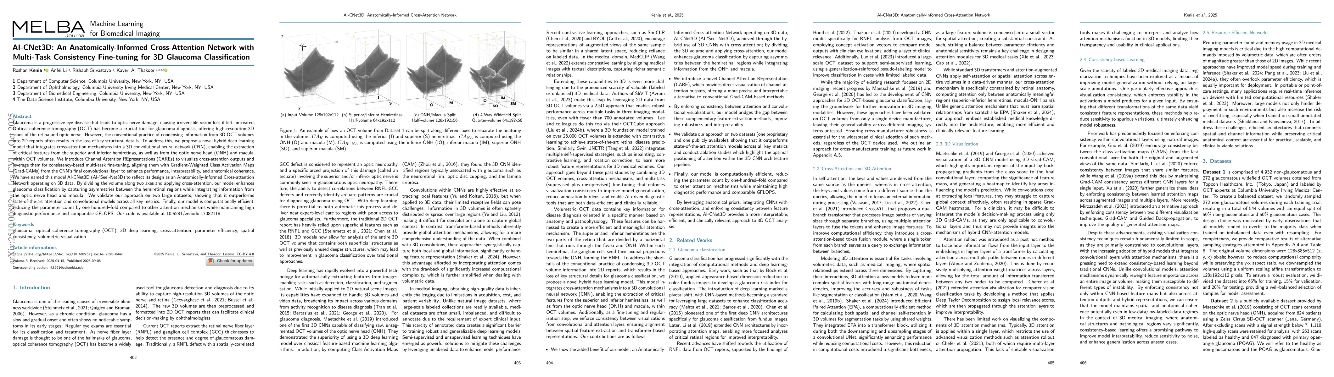
Glaucoma is a progressive eye disease that leads to optic nerve damage, causing irreversible vision loss if left untreated. Optical coherence tomography (OCT) has become a crucial tool for glaucoma diagnosis, offering high-resolution 3D scans of the retina and optic nerve. However, the conventional practice of condensing information from 3D OCT volumes into 2D reports often results in the loss of key structural details. To address this, we propose a novel hybrid deep learning model that integrates cross-attention mechanisms into a 3D convolutional neural network (CNN), enabling the extraction of critical features from the superior and inferior hemiretinas, as well as from the optic nerve head (ONH) and macula, within OCT volumes. We introduce Channel Attention REpresentations (CAREs) to visualize cross-attention outputs and leverage them for consistency-based multi-task fine-tuning, aligning them with Gradient-Weighted Class Activation Maps (Grad-CAMs) from the CNN's final convolutional layer to enhance performance, interpretability, and anatomical coherence. We have named this model AI-CNet3D (AI-`See'-Net3D) to reflect its design as an Anatomically-Informed Cross-attention Network operating on 3D data. By dividing the volume along two axes and applying cross-attention, our model enhances glaucoma classification by capturing asymmetries between the hemiretinal regions while integrating information from the optic nerve head and macula. We validate our approach on two large datasets, showing that it outperforms state-of-the-art attention and convolutional models across all key metrics. Finally, our model is computationally efficient, reducing the parameter count by one-hundred--fold compared to other attention mechanisms while maintaining high diagnostic performance and comparable GFLOPS.

Key Findings, in focus

Seven facets of this paper, analysed and brought into focus by AI.

AI-generated · verify against source Generated Oct 04, 2025
In focus · one-line synthesis

This research advances AI-driven ophthalmology by enabling more accurate and interpretable glaucoma detection through anatomically informed 3D analysis, which could improve early diagnosis and treatment outcomes.

01
MethodologyHow they did it

The study introduces a 3D cross-attention mechanism for widefield OCT analysis, combining anatomical priors with inter-region correlations within 3D volumes. It employs a hybrid CNN-attention model trained with joint unsupervised visualization consistency loss and BCE loss for fine-tuning.

02
ResultsWhat they found
  • The AI-CNet3D model outperforms traditional 3D CNNs and state-of-the-art spatial/channel attention methods in classification accuracy and computational efficiency
  • Superior-inferior and macula-nerve cross-attention mechanisms improve sensitivity, specificity, and overall performance for glaucoma detection
  • CARE visualizations highlight critical retinal structures like RNFL, GCC, and photoreceptors, enhancing model interpretability
03
SignificanceWhy it matters

This research advances AI-driven ophthalmology by enabling more accurate and interpretable glaucoma detection through anatomically informed 3D analysis, which could improve early diagnosis and treatment outcomes.

04
Technical ContributionsWhat's new technically

Proposes a novel 3D cross-attention framework that integrates anatomical relationships and inter-region correlations, along with a joint loss function for enhanced model interpretability and performance.

05
NoveltyWhat sets it apart

Introduces CARE (Cross-Attention Retinal Evaluation) technique for visualizing 3D attention mechanisms, providing deeper insights into retinal structure contributions compared to existing Grad-CAM methods.

06
LimitationsWhat to be cautious about
  • Zeiss visualization precision is lower due to lower resolution volumes
  • CARE and Grad-CAM consistency is stronger in later layers for true negatives but requires further validation
07
Future WorkWhere it goes next
  • Incorporate full datasets or external domain data for pre-training to improve robustness
  • Develop pseudo-segmentation masks for clinical validation and biomarker discovery
  • Explore applications in other ophthalmic diseases beyond glaucoma

Paper Details

Paper ID: 2510.00882
Comments: Accepted for publication at the Journal of Machine Learning for Biomedical Imaging (MELBA) https://melba-journal.org/2025:018
Categories:
cs.CV cs.LG

How to Cite This Paper

Reference manager: RIS EndNote

Related Papers

4 found


Discussion 0

This website uses cookies to ensure you get the best experience. Learn more