Publication
Metrics
AI Quick Summary
This study introduces an AI-driven image analysis system for segmenting and analyzing microbial cells using an automated pipeline that includes denoising, segmentation, post-processing, and quantitative feature extraction. The system enhances segmentation accuracy and enables efficient, high-resolution analysis of microbial growth and metabolism.
Paper Preview
Abstract
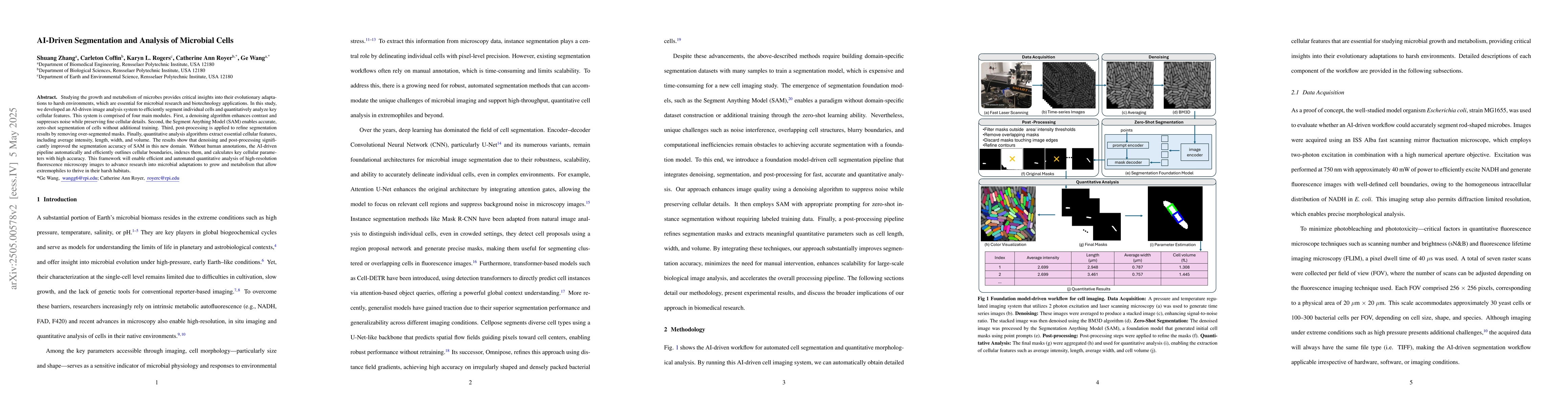
Studying the growth and metabolism of microbes provides critical insights into their evolutionary adaptations to harsh environments, which are essential for microbial research and biotechnology applications. In this study, we developed an AI-driven image analysis system to efficiently segment individual cells and quantitatively analyze key cellular features. This system is comprised of four main modules. First, a denoising algorithm enhances contrast and suppresses noise while preserving fine cellular details. Second, the Segment Anything Model (SAM) enables accurate, zero-shot segmentation of cells without additional training. Third, post-processing is applied to refine segmentation results by removing over-segmented masks. Finally, quantitative analysis algorithms extract essential cellular features, including average intensity, length, width, and volume. The results show that denoising and post-processing significantly improved the segmentation accuracy of SAM in this new domain. Without human annotations, the AI-driven pipeline automatically and efficiently outlines cellular boundaries, indexes them, and calculates key cellular parameters with high accuracy. This framework will enable efficient and automated quantitative analysis of high-resolution fluorescence microscopy images to advance research into microbial adaptations to grow and metabolism that allow extremophiles to thrive in their harsh habitats.
AI Key Findings
Get AI-generated insights about this paper's methodology, results, significance, and more — seven facets brought into focus.
Impact
Authors
PDF Preview
Citation Network
Current paper (gray), citations (green), references (blue)
Display is limited for performance on very large graphs.
Discussion 0