01
MethodologyHow they did it
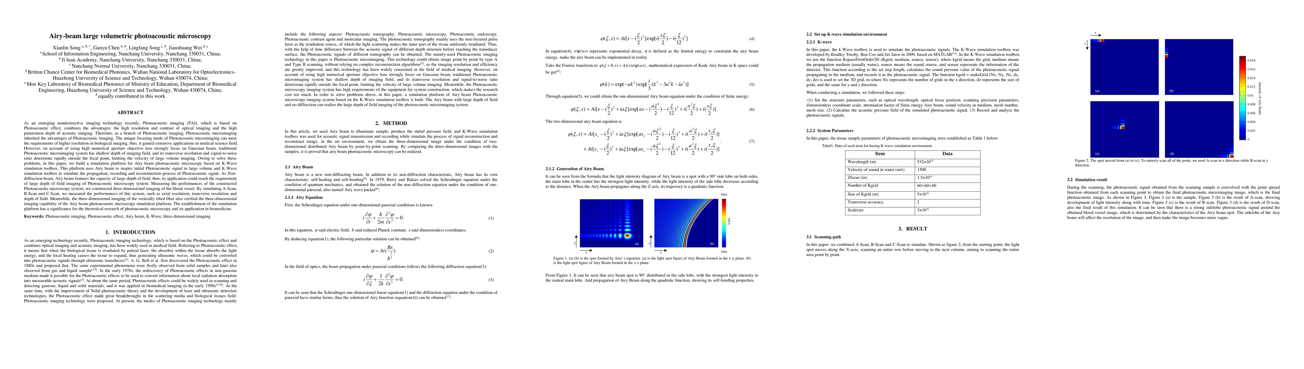
A simulation platform for Airy beam photoacoustic microscopy was built using the K-Wave simulation toolbox in MATLAB. The platform utilized Airy beam to illuminate the sample, producing an initial pressure field. The K-Wave toolbox was used for simulating acoustic signal transmission and recording, reconstructing signals, and obtaining three-dimensional images through point-by-point scanning in a two-dimensional distributed Airy beam environment.
Discussion 0