Algorithm-based diagnostic application for diabetic retinopathy detection
Publication
Metrics
AI Quick Summary
This paper presents an algorithm-based diagnostic application for automatic detection of diabetic retinopathy using ophthalmoscopic images and deep learning techniques. The application aims to improve early diagnosis efficiency and reduce diabetes-related visual impairment by analyzing retinal images and identifying characteristic lesions.
Paper Preview
Abstract
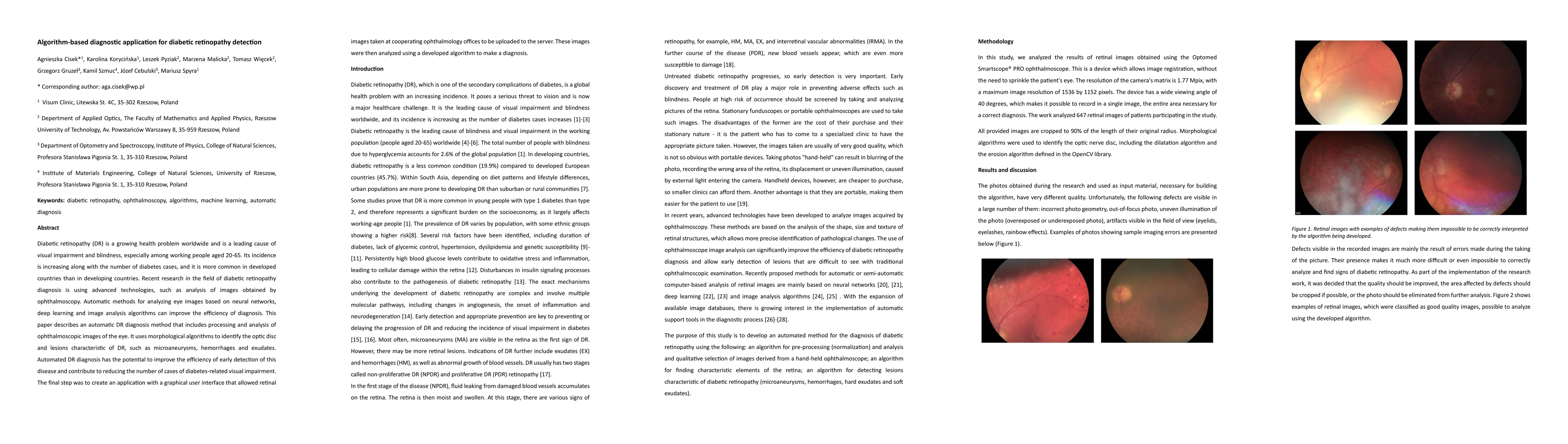
Diabetic retinopathy (DR) is a growing health problem worldwide and is a leading cause of visual impairment and blindness, especially among working people aged 20-65. Its incidence is increasing along with the number of diabetes cases, and it is more common in developed countries than in developing countries. Recent research in the field of diabetic retinopathy diagnosis is using advanced technologies, such as analysis of images obtained by ophthalmoscopy. Automatic methods for analyzing eye images based on neural networks, deep learning and image analysis algorithms can improve the efficiency of diagnosis. This paper describes an automatic DR diagnosis method that includes processing and analysis of ophthalmoscopic images of the eye. It uses morphological algorithms to identify the optic disc and lesions characteristic of DR, such as microaneurysms, hemorrhages and exudates. Automated DR diagnosis has the potential to improve the efficiency of early detection of this disease and contribute to reducing the number of cases of diabetes-related visual impairment. The final step was to create an application with a graphical user interface that allowed retinal images taken at cooperating ophthalmology offices to be uploaded to the server. These images were then analyzed using a developed algorithm to make a diagnosis.
AI Key Findings — Failed
Key findings generation failed. Failed to start generation process
Impact
Paper Details
Authors
PDF Preview
Key Terms
Citation Network
Current paper (gray), citations (green), references (blue)
Display is limited for performance on very large graphs.
Discussion 0