Publication
Metrics
Quick Actions
Paper Preview
Abstract
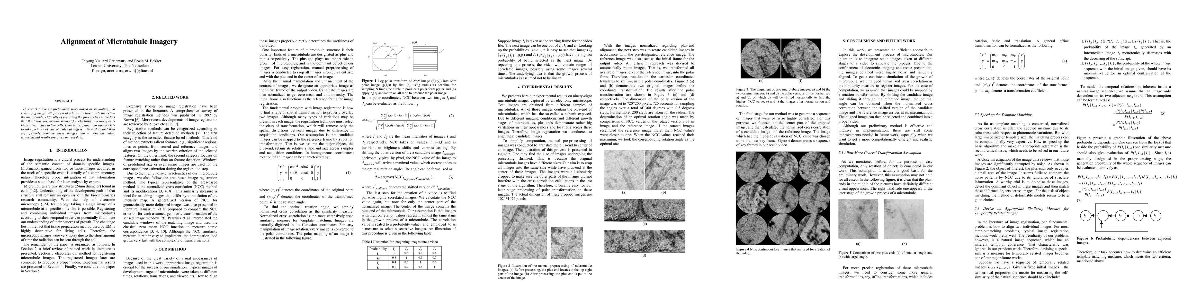
This work discusses preliminary work aimed at simulating and visualizing the growth process of a tiny structure inside the cell---the microtubule. Difficulty of recording the process lies in the fact that the tissue preparation method for electronic microscopes is highly destructive to live cells. Here in this paper, our approach is to take pictures of microtubules at different time slots and then appropriately combine these images into a coherent video. Experimental results are given on real data.
AI Key Findings
Get AI-generated insights about this paper's methodology, results, and significance.
Paper Details
How to Cite This Paper
@article{anon2011alignment,
  title         = {Alignment of Microtubule Imagery},
  year          = {2011},
  eprint        = {1105.6060},
  archivePrefix = {arXiv},
  primaryClass  = {cs.CV},
}(2011). Alignment of Microtubule Imagery. arXiv. https://arxiv.org/abs/1105.6060"Alignment of Microtubule Imagery." arXiv, 2011, arxiv.org/abs/1105.6060.PDF Preview
Key Terms
Citation Network
Current paper (gray), citations (green), references (blue)
Display is limited for performance on very large graphs.
Similar Papers
Found 4 papersActive Matter under Cyclic Stretch: Modeling Microtubule Alignment and Bundling
Takumi Tagaki, Seiya Nishikawa, Shuji Ishihara
Towards High-Resolution Alignment and Super-Resolution of Multi-Sensor Satellite Imagery
Philip Wootaek Shin, Vishal Gaur, Rahul Ramachandran et al.
Transferring Spatial Filters via Tangent Space Alignment in Motor Imagery BCIs
Tekin Gunasar, Virginia de Sa
No citations found for this paper.
Comments (0)