Altered structural connectivity of the left visual thalamus in developmental dyslexia

Source: ArXiv

AI Quick Summary

This paper reveals that individuals with developmental dyslexia exhibit reduced structural connectivity between the left visual thalamus (LGN) and the left middle temporal area V5/MT, but not between the left LGN and left primary visual cortex (V1). These findings suggest altered cortico-thalamic interactions may contribute to dyslexia symptoms.

Share: Share on X LinkedIn

Summary

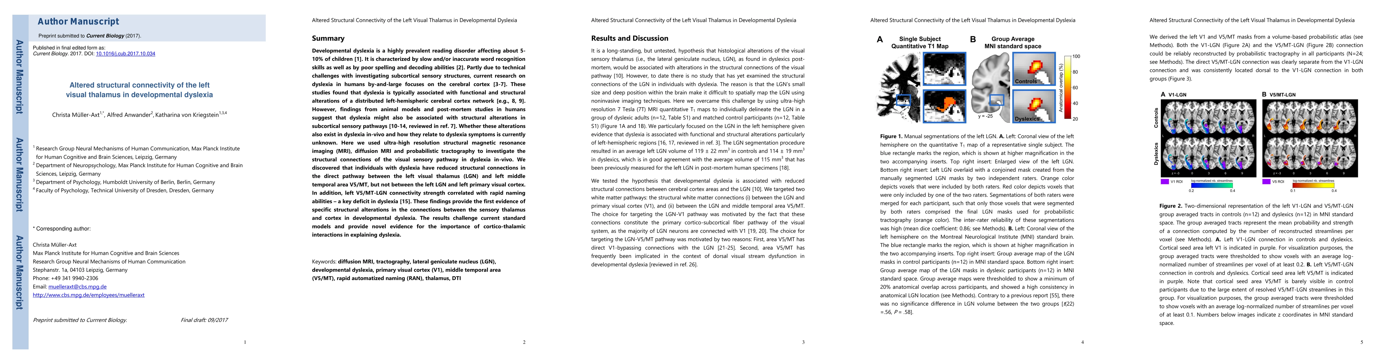
Developmental dyslexia is characterized by persistent reading and spelling deficits. Partly due to technical challenges with investigating subcortical sensory structures, current research on dyslexia in humans by-and-large focuses on the cerebral cortex. These studies found that dyslexia is typically associated with functional and structural alterations of a distributed left-hemispheric cerebral cortex network. However, findings from animal models and post-mortem studies in humans suggest that developmental dyslexia might also be associated with structural alterations in subcortical sensory pathways. Whether these alterations also exist in developmental dyslexia in-vivo and how they relate to dyslexia symptoms is currently unknown. Here we used ultra-high resolution structural magnetic resonance imaging (MRI), diffusion MRI and probabilistic tractography to investigate the structural connections of the visual sensory pathway in dyslexia in-vivo. We discovered that individuals with developmental dyslexia have reduced structural connections in the direct pathway between the left visual thalamus (LGN) and left middle temporal area V5/MT, but not between the left LGN and left primary visual cortex (V1). In addition, left V5/MT-LGN connectivity strength correlated with rapid naming abilities - a key deficit in dyslexia [14]. These findings provide the first evidence of specific structural alterations in the connections between the sensory thalamus and cortex in developmental dyslexia. The results challenge current standard models and provide novel evidence for the importance of cortico-thalamic interactions in explaining dyslexia.

AI Key Findings

Generated Sep 06, 2025

Methodology

Ultra-high resolution structural MRI, diffusion MRI, and probabilistic tractography were used to investigate the structural connections of the visual sensory pathway in dyslexia.

Key Results

  • Individuals with developmental dyslexia have reduced structural connections between the left visual thalamus (LGN) and left middle temporal area V5/MT.
  • Left V5/MT-LGN connectivity strength correlated with rapid naming abilities - a key deficit in dyslexia.
  • No significant alterations were found between the left LGN and left primary visual cortex (V1).

Significance

This study provides novel evidence for the importance of cortico-thalamic interactions in explaining dyslexia and challenges current standard models.

Technical Contribution

The use of probabilistic tractography to investigate structural connections in the visual sensory pathway.

Novelty

This study provides the first evidence of specific structural alterations in the connections between the sensory thalamus and cortex in developmental dyslexia.

Limitations

  • Technical challenges with investigating subcortical sensory structures may have limited the scope of this research.
  • The sample size was not explicitly stated, which may impact the generalizability of the findings.

Future Work

  • Investigating the neural mechanisms underlying dyslexia in other sensory pathways.
  • Examining the relationship between cortico-thalamic interactions and other cognitive deficits in dyslexia.

Paper Details

PDF Preview

Download PDF

Key Terms

left (0.303)
structural (0.301)
connections (0.246)
visual (0.224)
humans (0.212)
findings (0.165)
evidence (0.156)
ultra high (0.141)
Title Authors Year Actions

Comments (0)

This website uses cookies to ensure you get the best experience. Learn more