Publication
Metrics
AI Quick Summary
This research paper proposes a deep learning network for real-time segmentation of fluoroscopic images in total knee arthroplasty, achieving high accuracy with a mean-Average-Precision of 88.83% and a real-time speed of 20 frames per second, thus enhancing surgical planning and patient outcomes.
Paper Preview
Abstract
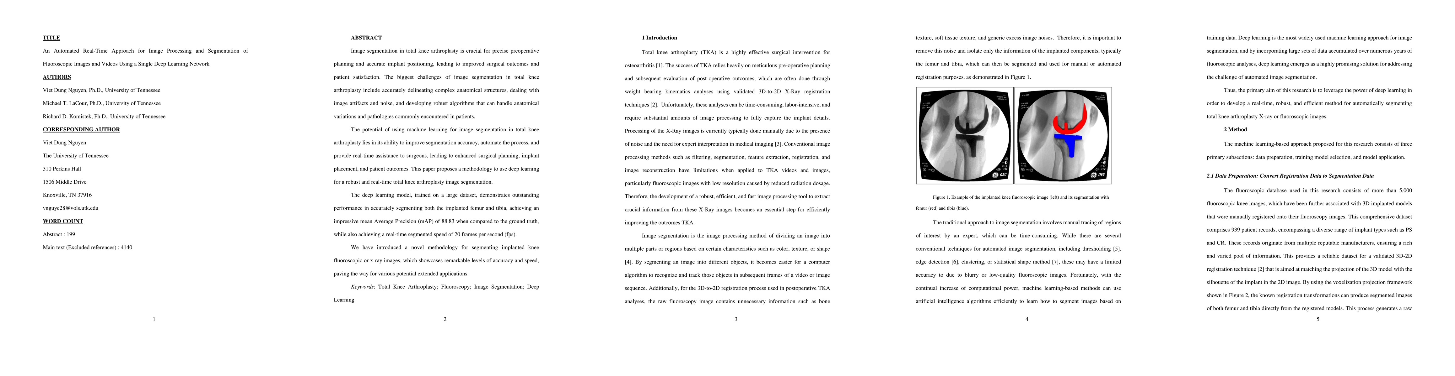
Image segmentation in total knee arthroplasty is crucial for precise preoperative planning and accurate implant positioning, leading to improved surgical outcomes and patient satisfaction. The biggest challenges of image segmentation in total knee arthroplasty include accurately delineating complex anatomical structures, dealing with image artifacts and noise, and developing robust algorithms that can handle anatomical variations and pathologies commonly encountered in patients. The potential of using machine learning for image segmentation in total knee arthroplasty lies in its ability to improve segmentation accuracy, automate the process, and provide real-time assistance to surgeons, leading to enhanced surgical planning, implant placement, and patient outcomes. This paper proposes a methodology to use deep learning for robust and real-time total knee arthroplasty image segmentation. The deep learning model, trained on a large dataset, demonstrates outstanding performance in accurately segmenting both the implanted femur and tibia, achieving an impressive mean-Average-Precision (mAP) of 88.83 when compared to the ground truth while also achieving a real-time segmented speed of 20 frames per second (fps). We have introduced a novel methodology for segmenting implanted knee fluoroscopic or x-ray images that showcases remarkable levels of accuracy and speed, paving the way for various potential extended applications.
AI Key Findings
Get AI-generated insights about this paper's methodology, results, significance, and more — seven facets brought into focus.
Impact
Paper Details
Authors
PDF Preview
Key Terms
Citation Network
Current paper (gray), citations (green), references (blue)
Display is limited for performance on very large graphs.
Discussion 0