ArxivLens

Quick Summary:

This paper presents an automated scanning transmission electron microscope (STEM) guided by sparse data analytics and AI, aiming to overcome current limitations in low-level instrument control and feature detection. The proposed closed-loop system uses machine learning to enable real-time experimental decision-making, facilitating high-throughput and statistical studies in material science.

DOI: 10.1017/S1431927622012065
arXiv Id: 2109.14772
Comments: 28 pages, 3 figures
Date Published: 2022-06-23
Date Updated: 2022-06-23

Key Terms (TF-IDF):

instrument (0.230)
instrument (0.229)
microscopy (0.225)
platform (0.218)
microscopy (0.217)
automated (0.214)
ai (0.213)
automated (0.210)
ai (0.207)
platform (0.206)
control (0.153)
control (0.153)
discrimination (0.148)
low level (0.148)
fly (0.146)
throughput (0.145)
quantum computing (0.145)
closed loop (0.144)
centralized (0.143)
informed (0.141)
License: http://arxiv.org/licenses/nonexclusive-distrib/1.0/

Summary:

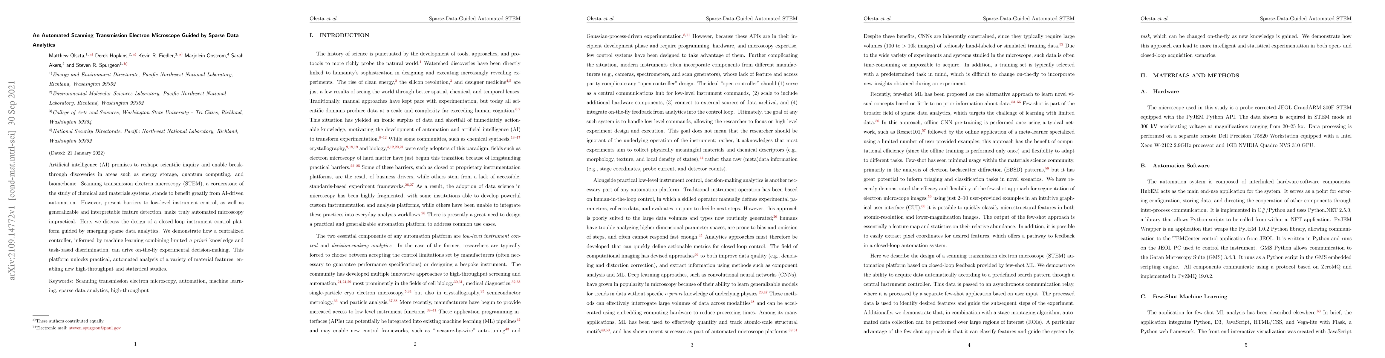
Artificial intelligence (AI) promises to reshape scientific inquiry and enable breakthrough discoveries in areas such as energy storage, quantum computing, and biomedicine. Scanning transmission electron microscopy (STEM), a cornerstone of the study of chemical and materials systems, stands to benefit greatly from AI-driven automation. However, present barriers to low-level instrument control, as well as generalizable and interpretable feature detection, make truly automated microscopy impractical. Here, we discuss the design of a closed-loop instrument control platform guided by emerging sparse data analytics. We demonstrate how a centralized controller, informed by machine learning combining limited $a$ $priori$ knowledge and task-based discrimination, can drive on-the-fly experimental decision-making. This platform unlocks practical, automated analysis of a variety of material features, enabling new high-throughput and statistical studies.

Citation & Reference Network Visualization

This graph shows how this paper is connected to other research. The current paper is shown in dark gray, papers that cite it are in green, and papers it references are in blue. You can:

Current Paper
Papers citing this work (Citations)
Papers cited by this work (References)

• Hover over any node to see the paper title

• Click a node to view that paper's details

• Drag nodes to rearrange the graph

• Use the zoom controls (bottom right) to zoom in/out

Citations and References

Title Authors Year Actions
Scientific exploration with expert knowledge (SEEK) in autonomous scanning probe microscopy with active learning
Utkarsh Pratiush, Hiroshi Funakubo, Rama K Vasudevan, Sergei V. Kalinin, Yongtao Liu
2024
View Paper
Unsupervised segmentation of irradiation-induced order-disorder phase transitions in electron microscopy
Arman Ter-Petrosyan, Jenna A. Bilbrey, Christina Doty, Bethany E. Matthews, Le Wang, Yingge Du, E. Lang, Khalid Hattar, S. Spurgeon
2023
View Paper
An artificial intelligence’s interpretation of complex high-resolution in situ transmission electron microscopy data
Xingzhi Wang, Chang Yan, Justin C. Ondry, Viraj Bodiwala, P. Ercius, A. P. Alivisatos
2023
View Paper
Statistical 3D morphology characterization of vaterite microspheres produced by engineered Escherichia coli.
A. Y. Lin, Zong-Yen Wu, Alexander J. Pattison, Isaak E. Müller, Yasuo Yoshikuni, Wolfgang Theis, P. Ercius
2023
View Paper
Dynamic STEM-EELS for single atom and defect measurement during electron beam transformations
Kevin M. Roccapriore, R. Torsi, Joshua Robinson, S. Kalinin, M. Ziatdinov
2023
View Paper
TEMGYM Advanced - NanoMi lens characterisation.
David Landers, I. Clancy, R. Dunin‐Borkowski, Dieter Weber, Andrew A. Stewart
2023
View Paper
Closed-loop optimization of nanoparticle synthesis enabled by robotics and machine learning
Jungwon Park, Young Min Kim, Seonghun Hong, Byungchan Han, Ki Tae Nam, Yousung Jung
2023
View Paper
Leveraging generative adversarial networks to create realistic scanning transmission electron microscopy images
Abid Khan, Chia-Hao Lee, Pinshane Y. Huang, B. Clark
2023
View Paper
Evaluating Stage Motion for Automated Electron Microscopy.
Kevin R Fiedler, M. Olszta, K. Yano, Christina Doty, Derek Hopkins, Sarah Akers, S. Spurgeon
2022
View Paper
Towards Augmented Microscopy with Reinforcement Learning-Enhanced Workflows
Michael Xu, Abinash Kumar, J. Lebeau
2022
View Paper
Probing Electron Beam Induced Transformations on a Single-Defect Level via Automated Scanning Transmission Electron Microscopy.
Kevin M. Roccapriore, M. Boebinger, O. Dyck, Ayana Ghosh, R. Unocic, S. Kalinin, M. Ziatdinov
2022
View Paper
Exploring Physics of Ferroelectric Domain Walls in Real Time: Deep Learning Enabled Scanning Probe Microscopy
Yongtao Liu, K. Kelley, H. Funakubo, S. Kalinin, M. Ziatdinov
2022
View Paper
Deep-learning-based prediction of nanoparticle phase transitions during in situ transmission electron microscopy
W. Fu, S. Spurgeon, Chongmin Wang, Yuyan Shao, Wei Wang, A. Peles
2022
View Paper
Using CycleGANs to Generate Realistic STEM Images for Machine Learning
Abid Khan, Chia-Hao Lee, Pinshane Y. Huang, B. Clark
2023
View Paper
Unsupervised and semi-supervised classification of order-disorder phase transitions in oxides
Arman Ter-Petrosyan, Jenna A. Bilbrey, Christina Doty, B. Matthews, Sarah Akers, S. Spurgeon
2023
View Paper

Comments (0)

This website uses cookies to ensure you get the best experience. Learn more