An efficient method to automate tooth identification and 3D bounding box extraction from Cone Beam CT Images
Publication
Metrics
AI Quick Summary
This research introduces an automated method for detecting and identifying teeth in Cone Beam CT images, utilizing a single-stage object detector for precise localization. The method also extracts 3D bounding boxes for each tooth, facilitating detailed dental analysis and integration into the Dentomo tool.
Paper Preview
Abstract
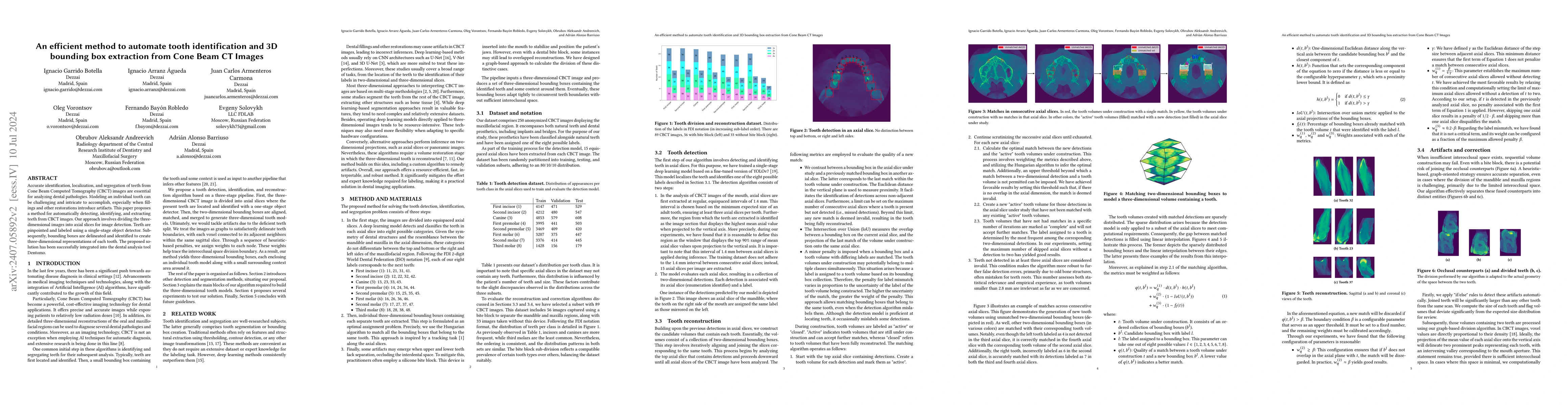
Accurate identification, localization, and segregation of teeth from Cone Beam Computed Tomography (CBCT) images are essential for analyzing dental pathologies. Modeling an individual tooth can be challenging and intricate to accomplish, especially when fillings and other restorations introduce artifacts. This paper proposes a method for automatically detecting, identifying, and extracting teeth from CBCT images. Our approach involves dividing the three-dimensional images into axial slices for image detection. Teeth are pinpointed and labeled using a single-stage object detector. Subsequently, bounding boxes are delineated and identified to create three-dimensional representations of each tooth. The proposed solution has been successfully integrated into the dental analysis tool Dentomo.
AI Key Findings
Get AI-generated insights about this paper's methodology, results, significance, and more — seven facets brought into focus.
Impact
Authors
PDF Preview
Key Terms
Citation Network
Current paper (gray), citations (green), references (blue)
Display is limited for performance on very large graphs.
Discussion 0