An Empirical Analysis for Zero-Shot Multi-Label Classification on COVID-19 CT Scans and Uncurated Reports
Publication
Metrics
AI Quick Summary
This research explores zero-shot multi-label classification for COVID-19 using CT scans and uncurated radiology reports, aiming to detect pulmonary embolisms and detailed lung features. The study evaluates various zero-shot models to address challenges in medical multimodal pretraining, offering insights for future advancements in medical image analysis.
Paper Preview
Abstract
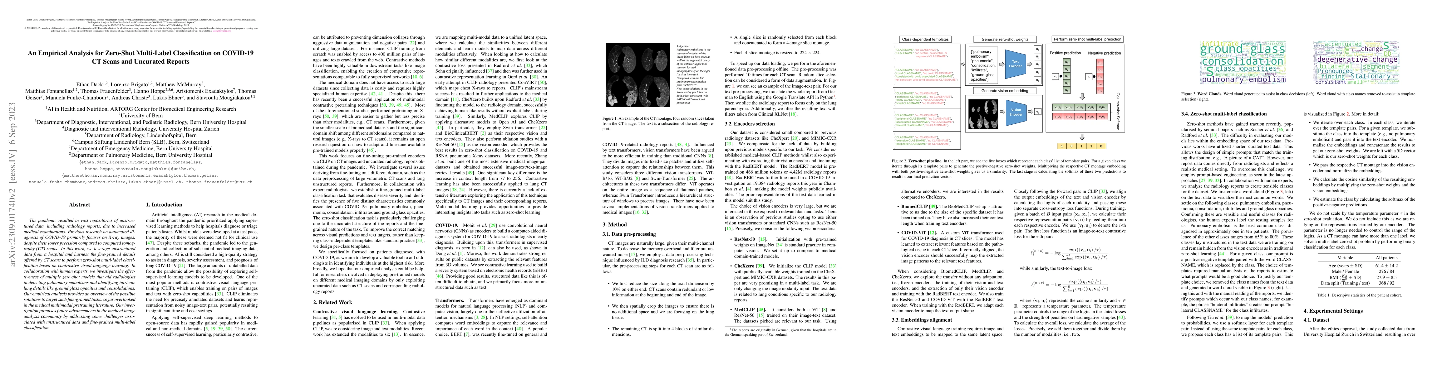
The pandemic resulted in vast repositories of unstructured data, including radiology reports, due to increased medical examinations. Previous research on automated diagnosis of COVID-19 primarily focuses on X-ray images, despite their lower precision compared to computed tomography (CT) scans. In this work, we leverage unstructured data from a hospital and harness the fine-grained details offered by CT scans to perform zero-shot multi-label classification based on contrastive visual language learning. In collaboration with human experts, we investigate the effectiveness of multiple zero-shot models that aid radiologists in detecting pulmonary embolisms and identifying intricate lung details like ground glass opacities and consolidations. Our empirical analysis provides an overview of the possible solutions to target such fine-grained tasks, so far overlooked in the medical multimodal pretraining literature. Our investigation promises future advancements in the medical image analysis community by addressing some challenges associated with unstructured data and fine-grained multi-label classification.
AI Key Findings
Get AI-generated insights about this paper's methodology, results, significance, and more — seven facets brought into focus.
Impact
Paper Details
Authors
PDF Preview
Key Terms
Citation Network
Current paper (gray), citations (green), references (blue)
Display is limited for performance on very large graphs.
Discussion 0