An explainable two-dimensional single model deep learning approach for Alzheimer's disease diagnosis and brain atrophy localization
Publication
Metrics
AI Quick Summary
This paper proposes a two-dimensional single model deep learning approach for diagnosing Alzheimer's disease and localizing brain atrophy using sMRI data. The method employs a suitable CNN model, a data augmentation strategy to mitigate overfitting, and Grad-CAM++ for explainable diagnosis, outperforming state-of-the-art methods and highlighting relevant brain regions.
Paper Preview
Abstract
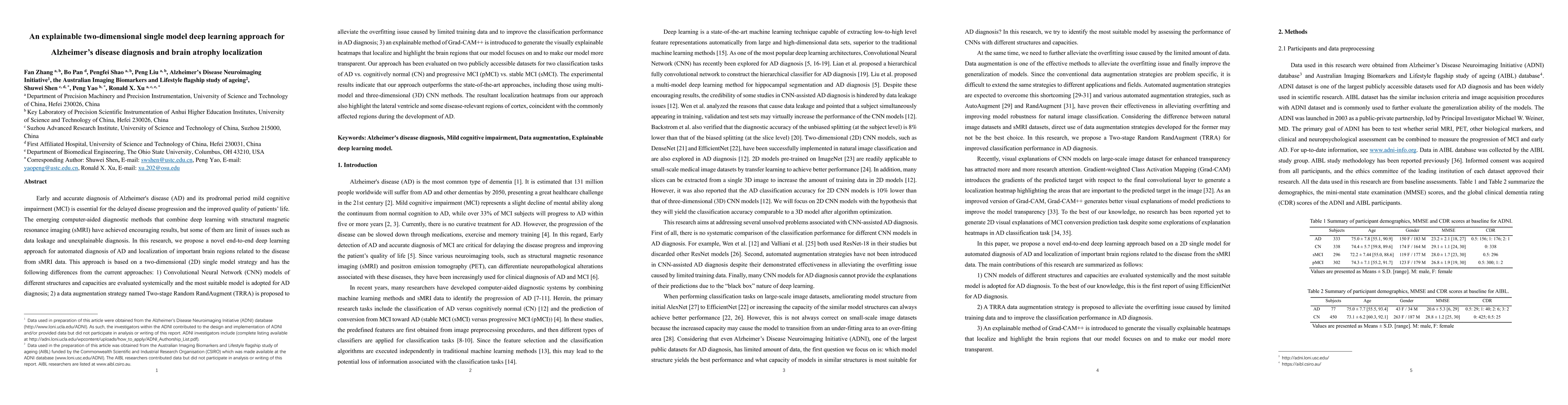
Early and accurate diagnosis of Alzheimer's disease (AD) and its prodromal period mild cognitive impairment (MCI) is essential for the delayed disease progression and the improved quality of patients'life. The emerging computer-aided diagnostic methods that combine deep learning with structural magnetic resonance imaging (sMRI) have achieved encouraging results, but some of them are limit of issues such as data leakage and unexplainable diagnosis. In this research, we propose a novel end-to-end deep learning approach for automated diagnosis of AD and localization of important brain regions related to the disease from sMRI data. This approach is based on a 2D single model strategy and has the following differences from the current approaches: 1) Convolutional Neural Network (CNN) models of different structures and capacities are evaluated systemically and the most suitable model is adopted for AD diagnosis; 2) a data augmentation strategy named Two-stage Random RandAugment (TRRA) is proposed to alleviate the overfitting issue caused by limited training data and to improve the classification performance in AD diagnosis; 3) an explainable method of Grad-CAM++ is introduced to generate the visually explainable heatmaps that localize and highlight the brain regions that our model focuses on and to make our model more transparent. Our approach has been evaluated on two publicly accessible datasets for two classification tasks of AD vs. cognitively normal (CN) and progressive MCI (pMCI) vs. stable MCI (sMCI). The experimental results indicate that our approach outperforms the state-of-the-art approaches, including those using multi-model and 3D CNN methods. The resultant localization heatmaps from our approach also highlight the lateral ventricle and some disease-relevant regions of cortex, coincident with the commonly affected regions during the development of AD.
AI Key Findings
Get AI-generated insights about this paper's methodology, results, significance, and more — seven facets brought into focus.
Impact
Paper Details
Authors
PDF Preview
Key Terms
Citation Network
Current paper (gray), citations (green), references (blue)
Display is limited for performance on very large graphs.
Discussion 0