An Exploration of 2D and 3D Deep Learning Techniques for Cardiac MR Image Segmentation
Publication
Metrics
AI Quick Summary
This paper presents a fully automated framework for segmenting cardiac MR images using 2D and 3D convolutional neural networks, achieving mean Dice coefficients of 0.950, 0.893, and 0.899 for the left ventricle, right ventricle, and myocardium respectively. The study finds that 2D networks are more effective due to the large slice thickness in cardiac MR images.
Paper Preview
Abstract
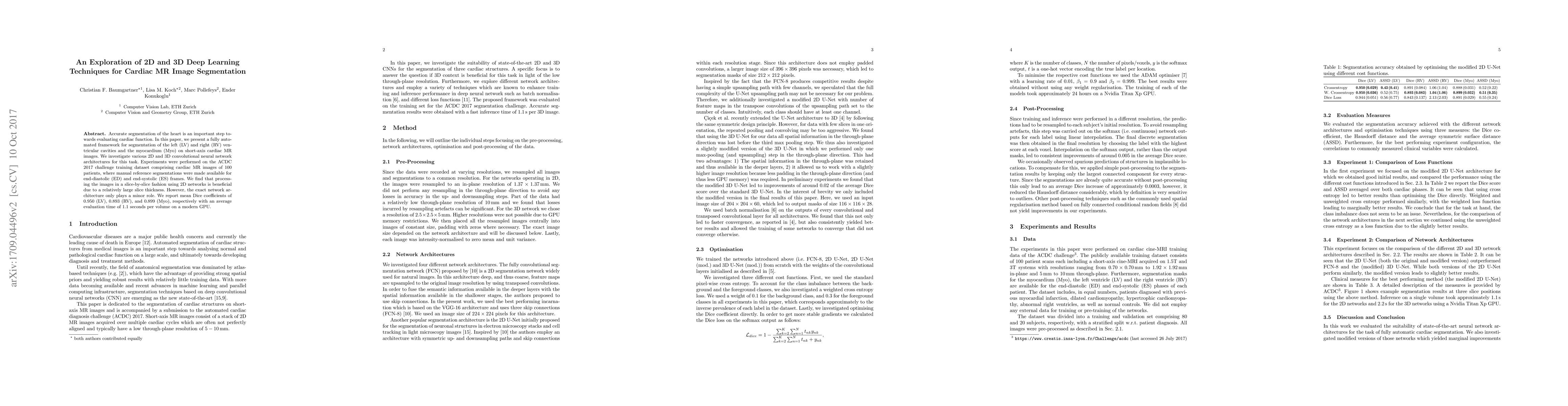
Accurate segmentation of the heart is an important step towards evaluating cardiac function. In this paper, we present a fully automated framework for segmentation of the left (LV) and right (RV) ventricular cavities and the myocardium (Myo) on short-axis cardiac MR images. We investigate various 2D and 3D convolutional neural network architectures for this task. We investigate the suitability of various state-of-the art 2D and 3D convolutional neural network architectures, as well as slight modifications thereof, for this task. Experiments were performed on the ACDC 2017 challenge training dataset comprising cardiac MR images of 100 patients, where manual reference segmentations were made available for end-diastolic (ED) and end-systolic (ES) frames. We find that processing the images in a slice-by-slice fashion using 2D networks is beneficial due to a relatively large slice thickness. However, the exact network architecture only plays a minor role. We report mean Dice coefficients of $0.950$ (LV), $0.893$ (RV), and $0.899$ (Myo), respectively with an average evaluation time of 1.1 seconds per volume on a modern GPU.
AI Key Findings
Get AI-generated insights about this paper's methodology, results, significance, and more — seven facets brought into focus.
Impact
Paper Details
PDF Preview
Key Terms
Citation Network
Current paper (gray), citations (green), references (blue)
Display is limited for performance on very large graphs.
Discussion 0