An Interactive Automation for Human Biliary Tree Diagnosis Using Computer Vision
Publication
Metrics
AI Quick Summary
This research develops an automated computer vision model to diagnose biliary tree conditions using MRI images, employing image processing to segment and extract features from the biliary tree. The model's accuracy in classifying normal and dilated bile ducts is demonstrated using a dataset from King Hussein Medical Center, showcasing a novel approach to biliary tree diagnosis.
Paper Preview
Abstract
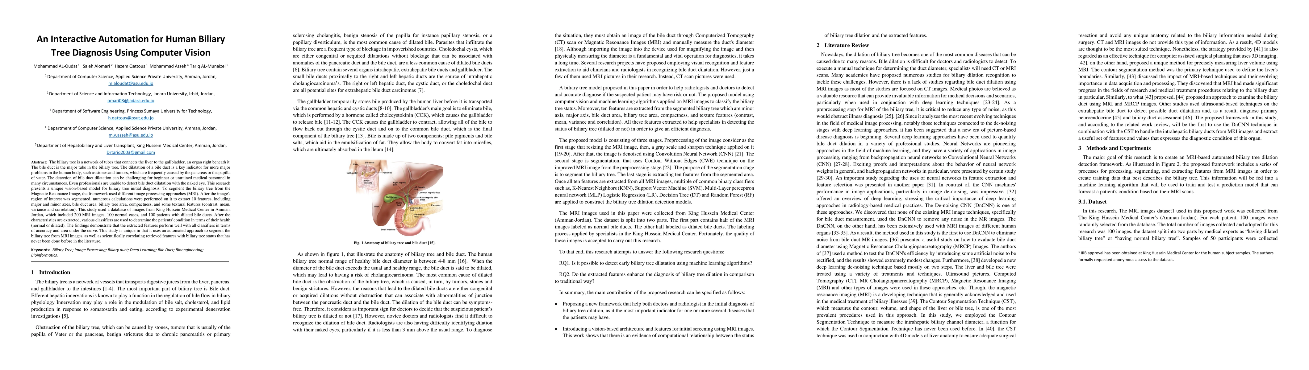
The biliary tree is a network of tubes that connects the liver to the gallbladder, an organ right beneath it. The bile duct is the major tube in the biliary tree. The dilatation of a bile duct is a key indicator for more major problems in the human body, such as stones and tumors, which are frequently caused by the pancreas or the papilla of vater. The detection of bile duct dilatation can be challenging for beginner or untrained medical personnel in many circumstances. Even professionals are unable to detect bile duct dilatation with the naked eye. This research presents a unique vision-based model for biliary tree initial diagnosis. To segment the biliary tree from the Magnetic Resonance Image, the framework used different image processing approaches (MRI). After the image's region of interest was segmented, numerous calculations were performed on it to extract 10 features, including major and minor axes, bile duct area, biliary tree area, compactness, and some textural features (contrast, mean, variance and correlation). This study used a database of images from King Hussein Medical Center in Amman, Jordan, which included 200 MRI images, 100 normal cases, and 100 patients with dilated bile ducts. After the characteristics are extracted, various classifiers are used to determine the patients' condition in terms of their health (normal or dilated). The findings demonstrate that the extracted features perform well with all classifiers in terms of accuracy and area under the curve. This study is unique in that it uses an automated approach to segment the biliary tree from MRI images, as well as scientifically correlating retrieved features with biliary tree status that has never been done before in the literature.
AI Key Findings
Get AI-generated insights about this paper's methodology, results, significance, and more — seven facets brought into focus.
Impact
Paper Details
Authors
PDF Preview
Key Terms
Citation Network
Current paper (gray), citations (green), references (blue)
Display is limited for performance on very large graphs.
Discussion 0