Summary
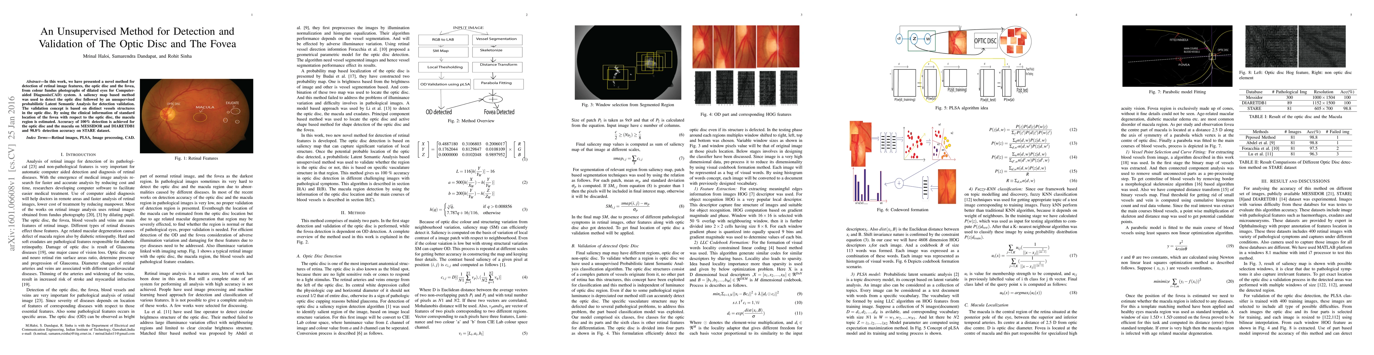
In this work, we have presented a novel method for detection of retinal image features, the optic disc and the fovea, from colour fundus photographs of dilated eyes for Computer-aided Diagnosis(CAD) system. A saliency map based method was used to detect the optic disc followed by an unsupervised probabilistic Latent Semantic Analysis for detection validation. The validation concept is based on distinct vessels structures in the optic disc. By using the clinical information of standard location of the fovea with respect to the optic disc, the macula region is estimated. Accuracy of 100\% detection is achieved for the optic disc and the macula on MESSIDOR and DIARETDB1 and 98.8\% detection accuracy on STARE dataset.
AI Key Findings
Get AI-generated insights about this paper's methodology, results, and significance.
Paper Details
PDF Preview
Key Terms
Citation Network
Current paper (gray), citations (green), references (blue)
Display is limited for performance on very large graphs.
Similar Papers
Found 4 papersJOINEDTrans: Prior Guided Multi-task Transformer for Joint Optic Disc/Cup Segmentation and Fovea Detection
Pujin Cheng, Xiaoying Tang, Li Lin et al.
JOINED : Prior Guided Multi-task Learning for Joint Optic Disc/Cup Segmentation and Fovea Detection
Xiaoying Tang, Li Lin, Zhiyuan Cai et al.
| Title | Authors | Year | Actions |
|---|
Comments (0)