Analysis of osteoporotic tissue using combination nonlinear optical imaging
Publication
Metrics
AI Quick Summary
This paper presents a novel method using combination nonlinear optical imaging techniques to analyze osteoporotic tissue without the need for paraffin removal, enabling non-destructive spectroscopic study. The approach successfully distinguishes osteoporotic from healthy bone using sum frequency generation and coherent anti-Stokes Raman scattering, with potential to validate past studies and minimize new sample production.
Paper Preview
Abstract
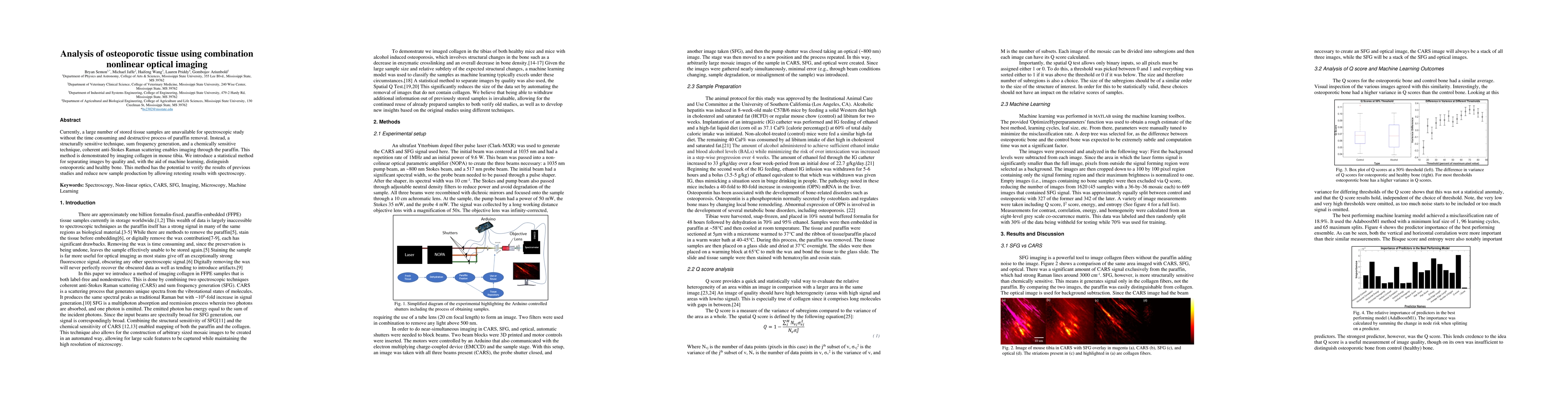
Currently, a large number of stored tissue samples are unavailable for spectroscopic study without the time consuming and destructive process of paraffin removal. Instead, a structurally sensitive technique, sum frequency generation, and a chemically sensitive technique, coherent anti-Stokes Raman scattering enables imaging through the paraffin. This method is demonstrated by imaging collagen in mouse tibia. We introduce a statistical method for separating images by quality and, with the aid of machine learning, distinguish osteoporotic and healthy bone. This method has the potential to verify the results of previous studies and reduce new sample production by allowing retesting results with spectroscopy.
AI Key Findings
Get AI-generated insights about this paper's methodology, results, significance, and more — seven facets brought into focus.
Impact
Paper Details
Authors
PDF Preview
Key Terms
Citation Network
Current paper (gray), citations (green), references (blue)
Display is limited for performance on very large graphs.
Discussion 0