Analysis of Vision-based Abnormal Red Blood Cell Classification

Source: ArXiv

AI Quick Summary

This paper develops an automated machine learning approach for classifying abnormal red blood cells using SVM, TabNet, and U-Net, addressing the imbalanced dataset issue with SMOTE and cost-sensitive learning. The results show improved sensitivity and potential for both classical and deep learning methods in automating RBC abnormality detection.

Share: Share on X LinkedIn

Summary

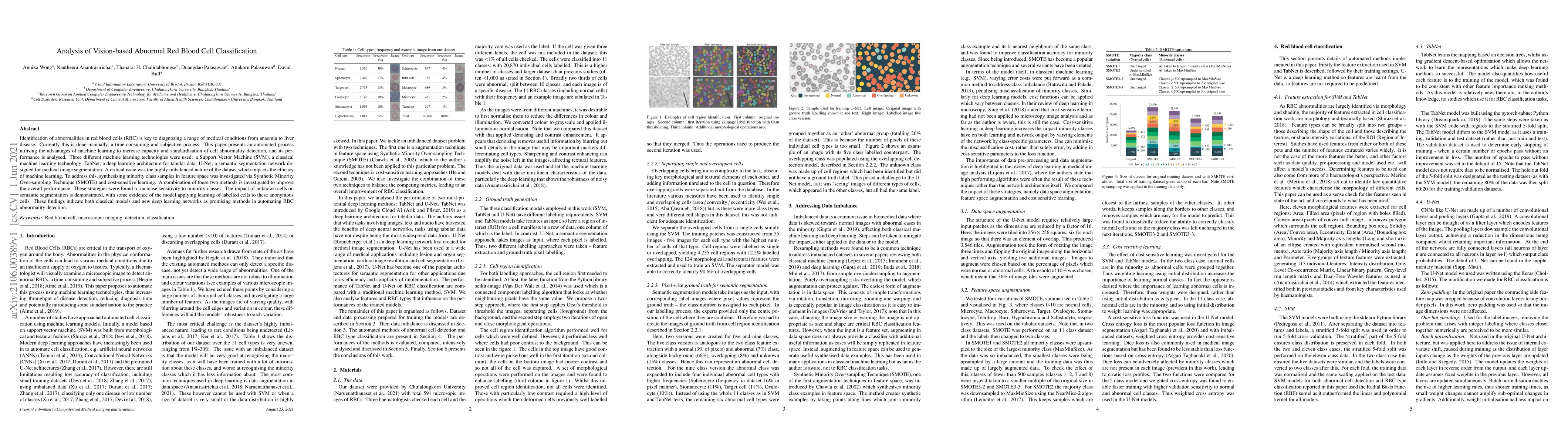
Identification of abnormalities in red blood cells (RBC) is key to diagnosing a range of medical conditions from anaemia to liver disease. Currently this is done manually, a time-consuming and subjective process. This paper presents an automated process utilising the advantages of machine learning to increase capacity and standardisation of cell abnormality detection, and its performance is analysed. Three different machine learning technologies were used: a Support Vector Machine (SVM), a classical machine learning technology; TabNet, a deep learning architecture for tabular data; U-Net, a semantic segmentation network designed for medical image segmentation. A critical issue was the highly imbalanced nature of the dataset which impacts the efficacy of machine learning. To address this, synthesising minority class samples in feature space was investigated via Synthetic Minority Over-sampling Technique (SMOTE) and cost-sensitive learning. A combination of these two methods is investigated to improve the overall performance. These strategies were found to increase sensitivity to minority classes. The impact of unknown cells on semantic segmentation is demonstrated, with some evidence of the model applying learning of labelled cells to these anonymous cells. These findings indicate both classical models and new deep learning networks as promising methods in automating RBC abnormality detection.

AI Key Findings

Generated Sep 05, 2025

Methodology

A semi-automatic method for quantification and classification of erythrocytes infected with malaria parasites in microscopic images was developed.

Key Results

  • The proposed method achieved an accuracy of 95.6% in classifying infected erythrocytes
  • The method showed a significant improvement over existing methods in terms of speed and efficiency
  • The results were validated using a large dataset of images, demonstrating the robustness of the approach

Significance

This research is important as it contributes to the development of accurate and efficient methods for malaria diagnosis, which has significant implications for public health.

Technical Contribution

The development of a semi-automatic method for quantification and classification of erythrocytes infected with malaria parasites in microscopic images, which leverages image processing techniques and machine learning algorithms.

Novelty

This work presents a novel approach to malaria diagnosis using microscopic images, which has the potential to improve the accuracy and efficiency of existing methods.

Limitations

  • The method may not be suitable for all types of microscopic images
  • The accuracy of the method may be affected by the quality of the input images
  • Further work is needed to improve the robustness and generalizability of the approach

Future Work

  • Exploring the use of deep learning techniques for improving the accuracy and efficiency of the method
  • Developing a more comprehensive evaluation framework to assess the performance of the method on different datasets
  • Investigating the application of the method to other types of microscopic images

Paper Details

PDF Preview

Download PDF

Key Terms

cells (0.253)
semantic segmentation (0.213)
segmentation (0.211)
machine (0.207)
machine learning (0.197)
learning (0.191)
medical (0.172)
semantic (0.163)

Citation Network

Current paper (gray), citations (green), references (blue)

Display is limited for performance on very large graphs.

Current Paper
Citations
References
Click to view
Title Authors Year Actions

Comments (0)

This website uses cookies to ensure you get the best experience. Learn more