Quick Summary:

Analytic Fourier Ptychotomography (AFP) presents a novel computational microscopy technique for high-resolution, aberration-free 3D refractive index imaging without iterative optimization. AFP enhances image quality and resolution by correcting optical aberrations and extending the synthetic numerical aperture, offering significant improvements over existing methods.

Summary

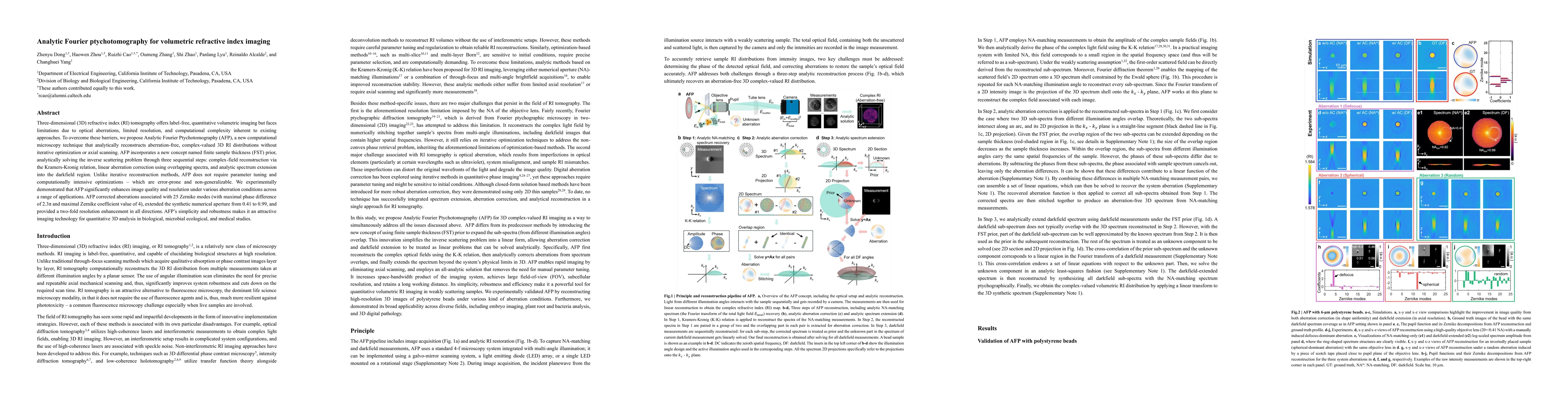
Three-dimensional (3D) refractive index (RI) tomography offers label-free, quantitative volumetric imaging but faces limitations due to optical aberrations, limited resolution, and the computational complexity inherent to existing approaches. To overcome these barriers, we propose Analytic Fourier Ptychotomography (AFP), a new computational microscopy technique that analytically reconstructs aberration-free, complex-valued 3D RI distributions without iterative optimization or axial scanning. AFP incorporates a new concept called the finite sample thickness (FST) prior, and analytically solves the inverse scattering problem through three sequential steps: complex-field reconstruction via the Kramers-Kronig relation, linear aberration correction using overlapping spectra, and analytic spectrum extension into the darkfield region. Unlike iterative reconstruction methods, AFP does not require parameter tuning or computationally intensive optimizations, which are often error-prone and non-generalizable. We experimentally demonstrate that AFP significantly enhances image quality and resolution under various aberration conditions across a range of applications. AFP corrected aberrations associated with 25 Zernike modes (with a maximal phase difference of 2.3$\pi$ and maximal Zernike coefficient value of 4), extended the synthetic numerical aperture from 0.41 to 0.99, and provided a two-fold resolution enhancement in all directions. AFP's simplicity and robustness make it an attractive imaging technology for quantitative 3D analysis in biological, microbial ecological, and medical studies.

AI Key Findings

Generated Jun 09, 2025

Methodology

The research introduces Analytic Fourier Ptychotomography (AFP), a new computational microscopy technique that analytically reconstructs aberration-free, complex-valued 3D refractive index distributions without iterative optimization or axial scanning. AFP uses three sequential steps: complex-field reconstruction via the Kramers-Kronig relation, linear aberration correction using overlapping spectra, and analytic spectrum extension into the darkfield region.

Key Results

  • AFP significantly enhances image quality and resolution under various aberration conditions.
  • AFP corrected aberrations associated with 25 Zernike modes, extended the synthetic numerical aperture from 0.41 to 0.99, and provided a two-fold resolution enhancement in all directions.
  • AFP was experimentally validated using polystyrene beads, mouse embryos, wheat roots, and human gastric tissues.
  • AFP demonstrated label-free volumetric imaging capabilities for biological, microbial ecological, and medical studies.

Significance

This research is significant as it presents a novel, analytic, and robust computational microscopy technique (AFP) for 3D refractive index tomography, overcoming limitations of existing iterative methods. AFP's simplicity and generalizability make it an attractive technology for quantitative 3D analysis across various fields, including biology, medicine, and plant science.

Technical Contribution

AFP's analytic formulation eliminates the need for parameter tuning, making it highly robust and generalizable across different sample types and system configurations. It extends the resolution beyond the NA of the objective lens through linear 3D spectrum extension, while preserving long working distances when using low-magnification objective lenses.

Novelty

AFP stands out due to its analytic approach, which contrasts with existing iterative reconstruction methods. Its ability to correct aberrations and extend resolution without optimization or iterative processes is a novel contribution to computational microscopy.

Limitations

  • AFP has been validated only in the weakly scattering regime, where the scattered light intensity from the sample is less than 25% of the incident light intensity.
  • Currently, AFP does not account for multiple scattering effects in optically thick samples or those with significant RI differences relative to the background.

Future Work

  • Explore AFP's potential for live cell and organoid imaging applications.
  • Investigate the integration of AFP with expansion microscopy for imaging cellular contents distantly embedded from the coverslip.

Paper Details

PDF Preview

Download PDF

Key Terms

No key terms available for this paper.

No citations found for this paper.

Comments (0)

This website uses cookies to ensure you get the best experience. Learn more